
Page 698 - Textbook of Pathology, 6th Edition
P. 698
682 in the urinary tract, and in debilitated or immunosuppressed
patients.
MORPHOLOGIC FEATURES. Grossly, well-developed
cases of acute pyelonephritis show enlarged and swollen
kidney that bulges on section. The cut surface shows small,
yellow-white abscesses with a haemorrhagic rim. These
abscesses may be several millimetres across and are
situated mainly in the cortex.
Microscopically, acute pyelonephritis is characterised by
extensive acute inflammation involving the interstitium
and causing destruction of the tubules. Generally, the
glomeruli and renal blood vessels show considerable
resistance to infection and are spared. The acute
inflammation may be in the form of large number of
neutrophils in the interstitial tissue and bursting into
tubules, or may form focal neutrophilic abscesses in the
renal parenchyma.
CLINICAL FEATURES. Classically, acute pyelonephritis has
an acute onset with chills, fever, loin pain, lumbar tenderness, Figure 22.30 Pyonephrosis. The kidney is enlarged and has
dysuria and frequency of micturition. Urine will show increased perinephric fat in the hilum. Sectioned surface shows markedly
dilated pelvis and calyces having irregular and ragged inner surface and
bacteria in excess of 100,000/ml, pus cells and pus cell casts containing necrotic debris and pus.
in the urinary sediment. Institution of specific antibiotics,
after identification of bacteria by culture followed by
sensitivity test, eradicates the infection in majority of patients. bed—reflux nephropathy and obstructive pyelonephritis
(Fig. 22.31):
COMPLICATIONS. Complications of acute pyelonephritis 1. Reflux nephropathy. Reflux of urine from the bladder
are encountered more often in patients with diabetes mellitus into one or both the ureters during micturition is the major
or with urinary tract obstruction. Following are the three cause of chronic pyelonephritis. Vesicoureteric reflux is
SECTION III
important complications of acute pyelonephritis:
particularly common in children, especially in girls, due to
1. Papillary necrosis. Papillary necrosis or necrotising congenital absence or shortening of the intravesical portion
papillitis develops more commonly in analgesic abuse of the ureter so that ureter is not compressed during the act
nephropathy and in sickle cell disease but may occur as a of micturition. Reflux results in increase in pressure in the
complication of acute pyelonephritis as well. It may affect renal pelvis so that the urine is forced into renal tubules which
one or both kidneys. is eventually followed by damage to the kidney and scar
formation (Fig. 22.29). Vesicoureteric reflux is more common
Grossly, the necrotic papillae are yellow to grey-white, in patients with urinary tract infection, whether symptomatic
sharply-defined areas with congested border and resemble or asymptomatic, but reflux of sterile urine can also cause
infarction. The pelvis may be dilated. renal damage.
Microscopically, necrotic tissue is separated from the
Systemic Pathology
viable tissue by a dense zone of polymorphs. The necrotic 2. Obstructive pyelonephritis. Obstruction to the outflow
area shows characteristic coagulative necrosis as seen in of urine at different levels predisposes the kidney to infection
renal infarcts. (page 690). Recurrent episodes of such obstruction and
infection result in renal damage and scarring. Rarely,
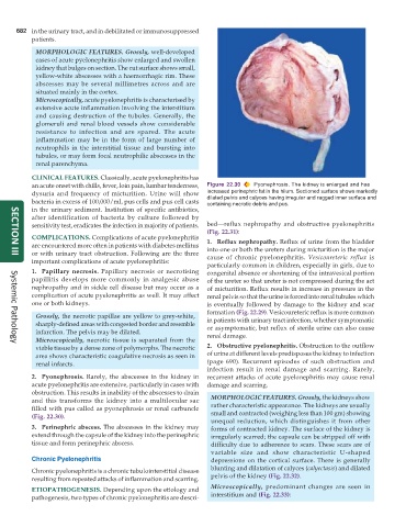
2. Pyonephrosis. Rarely, the abscesses in the kidney in recurrent attacks of acute pyelonephritis may cause renal
acute pyelonephritis are extensive, particularly in cases with damage and scarring.
obstruction. This results in inability of the abscesses to drain
and this transforms the kidney into a multilocular sac MORPHOLOGIC FEATURES. Grossly, the kidneys show
filled with pus called as pyonephrosis or renal carbuncle rather characteristic appearance. The kidneys are usually
(Fig. 22.30). small and contracted (weighing less than 100 gm) showing
unequal reduction, which distinguishes it from other
3. Perinephric abscess. The abscesses in the kidney may forms of contracted kidney. The surface of the kidney is
extend through the capsule of the kidney into the perinephric irregularly scarred; the capsule can be stripped off with
tissue and form perinephric abscess. difficulty due to adherence to scars. These scars are of
variable size and show characteristic U-shaped
Chronic Pyelonephritis depressions on the cortical surface. There is generally
Chronic pyelonephritis is a chronic tubulointerstitial disease blunting and dilatation of calyces (calyectasis) and dilated
resulting from repeated attacks of inflammation and scarring. pelvis of the kidney (Fig. 22.32).
Microscopically, predominant changes are seen in
ETIOPATHOGENESIS. Depending upon the etiology and
pathogenesis, two types of chronic pyelonephritis are descri- interstitium and (Fig. 22.33):