
Page 699 - Textbook of Pathology, 6th Edition
P. 699
683
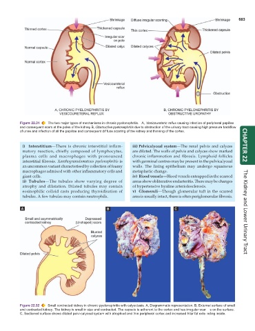
Figure 22.31 The two major types of mechanisms in chronic pyelonephritis. A, Vesicoureteric reflux causing infection of peripheral papillae
and consequent scars at the poles of the kidney. B, Obstructive pyelonephritis due to obstruction of the urinary tract causing high pressure backflow
of urine and infection of all the papillae and consequent dif fuse scarring of the kidney and thinning of the cortex.
i) Interstitium—There is chronic interstitial inflam- iii) Pelvicalyceal system—The renal pelvis and calyces CHAPTER 22
matory reaction, chiefly composed of lymphocytes, are dilated. The walls of pelvis and calyces show marked
plasma cells and macrophages with pronounced chronic inflammation and fibrosis. Lymphoid follicles
interstitial fibrosis. Xanthogranulomatous pyelonephritis is with germinal centres may be present in the pelvicalyceal
an uncommon variant characterised by collection of foamy walls. The lining epithelium may undergo squamous
macrophages admixed with other inflammatory cells and metaplastic change.
giant cells. iv) Blood vessels—Blood vessels entrapped in the scarred
ii) Tubules—The tubules show varying degree of areas show obliterative endarteritis. There may be changes
atrophy and dilatation. Dilated tubules may contain of hypertensive hyaline arteriolosclerosis.
eosinophilic colloid casts producing thyroidisation of v) Glomeruli—Though glomerular tuft in the scarred
tubules. A few tubules may contain neutrophils. area is usually intact, there is often periglomerular fibrosis. The Kidney and Lower Urinary Tract
Figure 22.32 Small contracted kidney in chronic pyelonephritis with calyectasis. A, Diagrammatic representation. B, External surface of small
and contracted kidney. The kidney is small in size and contracted. The capsule is adherent to the cortex and has irregular scar s on the surface.
C, Sectioned surface shows dilated pelvi-calyceal system with atrophied and thin peripheral cortex and increased hilar fat exte nding inside.