


Page 71 - Textbook of Pathology, 6th Edition
P. 71
ATROPHY                                                                                                  55
           Reduction of the number and size of parenchymal cells of an
           organ or its parts which was once normal is called atrophy
           (compared from hypoplasia which is the term used for
           developmentally small size, and aplasia for extreme failure
           of development so that only rudimentary tissue is present).                                                CHAPTER 3
           CAUSES. Atrophy may occur from physiologic or pathologic
           causes:
           A. Physiologic atrophy.  Atrophy is a normal process of
           aging in some tissues, which could be due to loss of endocrine
           stimulation or arteriosclerosis. For example:
           i) Atrophy of lymphoid tissue in lymph nodes, appendix
           and thymus.
           ii) Atrophy of gonads after menopause.
           iii) Atrophy of brain with aging.
           B. Pathologic atrophy. The causes are as under:
           1. Starvation atrophy. In starvation, there is first depletion
           of carbohydrate and fat stores followed by protein                                                         Cell Injury and Cellular Adaptations
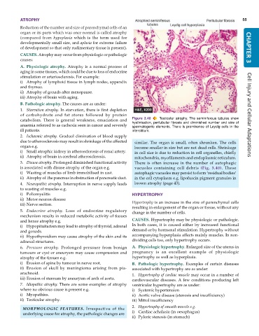
           catabolism. There is general weakness, emaciation and  Figure 3.40  Testicular atrophy. The seminiferous tubules show
           anaemia referred to as cachexia seen in cancer and severely  hyalinisation, peritubular fibrosis and diminished number and size of
                                                               spermatogenic elements. There is prominence of Leydig cells in the
           ill patients.                                       interstitium.
           2. Ischaemic atrophy. Gradual diminution of blood supply
           due to atherosclerosis may result in shrinkage of the affected  similar. The organ is small, often shrunken. The cells
           organ e.g.                                            become smaller in size but are not dead cells. Shrinkage
           i) Small atrophic kidney in atherosclerosis of renal artery.  in cell size is due to reduction in cell organelles, chiefly
           ii) Atrophy of brain in cerebral atherosclerosis.     mitochondria, myofilaments and endoplasmic reticulum.
           3. Disuse atrophy. Prolonged diminished functional activity  There is often increase in the number of autophagic
           is associated with disuse atrophy of the organ e.g.   vacuoles containing cell debris  (Fig.  3.40). These
           i) Wasting of muscles of limb immobilised in cast.    autophagic vacuoles may persist to form ‘residual bodies’
           ii) Atrophy of .the pancreas in obstruction of pancreatic duct.  in the cell cytoplasm e.g. lipofuscin pigment granules in
           4. Neuropathic atrophy. Interruption in nerve supply leads  brown atrophy (page 43).
           to wasting of muscles e.g.
           i) Poliomyelitis                                    HYPERTROPHY
           ii) Motor neuron disease
           iii) Nerve section.                                 Hypertrophy is an increase in the size of parenchymal cells
                                                               resulting in enlargement of the organ or tissue, without any
           5. Endocrine atrophy. Loss of endocrine regulatory  change in the number of cells.
           mechanism results in reduced metabolic activity of tissues
           and hence atrophy e.g.                              CAUSES. Hypertrophy may be physiologic or pathologic.
           i) Hypopituitarism may lead to atrophy of thyroid, adrenal  In both cases, it is caused either by increased functional
           and gonads.                                         demand or by hormonal stimulation. Hypertrophy without
           ii) Hypothyroidism may cause atrophy of the skin and its  accompanying hyperplasia affects mainly muscles. In non-
           adnexal structures.                                 dividing cells too, only hypertrophy occurs.
           6. Pressure atrophy. Prolonged pressure from benign  A. Physiologic hypertrophy. Enlarged size of the uterus in
           tumours or cyst or aneurysm may cause compression and  pregnancy is an excellent example of physiologic
           atrophy of the tissues e.g.                         hypertrophy as well as hyperplasia.
           i) Erosion of spine by tumour in nerve root.        B. Pathologic hypertrophy.  Examples of certain diseases
           ii) Erosion of skull by meningioma arising from pia-  associated with hypertrophy are as under:
           arachnoid.                                          1. Hypertrophy of cardiac muscle may occur in a number of
           iii) Erosion of sternum by aneurysm of arch of aorta.  cardiovascular diseases. A few conditions producing left
           7. Idiopathic atrophy. There are some examples of atrophy  ventricular hypertrophy are as under:
           where no obvious cause is present e.g.              i) Systemic hypertension
           i) Myopathies.                                      ii) Aortic valve disease (stenosis and insufficiency)
           ii) Testicular atrophy.                             iii) Mitral insufficiency
            MORPHOLOGIC FEATURES. Irrespective of the          2. Hypertrophy of smooth muscle e.g.
            underlying cause for atrophy, the pathologic changes are  i) Cardiac achalasia (in oesophagus)
                                                               ii) Pyloric stenosis (in stomach)

