


Page 68 - Textbook of Pathology, 6th Edition
P. 68
52


     SECTION I


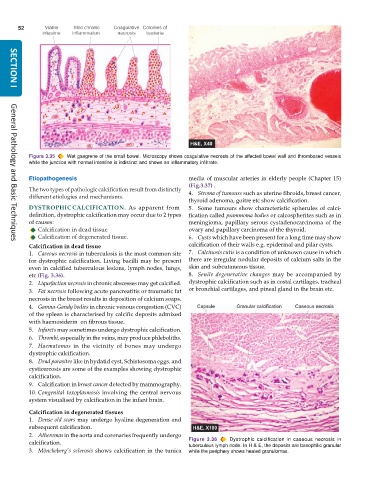
           Figure 3.35  Wet gangrene of the small bowel. Microscopy shows coagulative necrosis of the affected bowel wall and thrombosed vessels
           while the junction with normal intestine is indistinct and shows an inflammatory infiltrate.

           Etiopathogenesis                                    media of muscular arteries in elderly people (Chapter 15)
                                                               (Fig.3.37) .
           The two types of pathologic calcification result from distinctly  4. Stroma of tumours such as uterine fibroids, breast cancer,
           different etiologies and mechanisms.
                                                               thyroid adenoma, goitre etc show calcification.
           DYSTROPHIC CALCIFICATION.  As apparent from         5. Some tumours show characteristic spherules of calci-
           definition, dystrophic calcification may occur due to 2 types  fication called psammoma bodies or calcospherites such as in
           of causes:                                          meningioma, papillary serous cystadenocarcinoma of the
              Calcification in dead tissue                     ovary and papillary carcinoma of the thyroid.
              Calcification of degenerated tissue.             6. Cysts which have been present for a long time may show
     General Pathology and Basic Techniques
           Calcification in dead tissue                        calcification of their walls e.g. epidermal and pilar cysts.
           1. Caseous necrosis in tuberculosis is the most common site  7. Calcinosis cutis is a condition of unknown cause in which
           for dystrophic calcification. Living bacilli may be present  there are irregular nodular deposits of calcium salts in the
           even in calcified tuberculous lesions, lymph nodes, lungs,  skin and subcutaneous tissue.
           etc (Fig. 3.36).                                    8. Senile degenerative changes may be accompanied by
           2. Liquefaction necrosis in chronic abscesses may get calcified.  dystrophic calcification such as in costal cartilages, tracheal
           3. Fat necrosis following acute pancreatitis or traumatic fat  or bronchial cartilages, and pineal gland in the brain etc.
           necrosis in the breast results in deposition of calcium soaps.
           4. Gamna-Gandy bodies in chronic venous congestion (CVC)
           of the spleen is characterised by calcific deposits admixed
           with haemosiderin  on fibrous tissue.
           5. Infarcts may sometimes undergo dystrophic calcification.
           6. Thrombi, especially in the veins, may produce phleboliths.
           7. Haematomas in the vicinity of bones may undergo
           dystrophic calcification.
           8. Dead parasites like in hydatid cyst, Schistosoma eggs, and
           cysticercosis are some of the examples showing dystrophic
           calcification.
           9. Calcification in breast cancer detected by mammography.
           10. Congenital toxoplasmosis involving the central nervous
           system visualised by calcification in the infant brain.
           Calcification in degenerated tissues
           1. Dense old scars may undergo hyaline degeneration and
           subsequent calcification.
           2. Atheromas in the aorta and coronaries frequently undergo
           calcification.                                      Figure 3.36   Dystrophic calcification in caseous necrosis in
                                                               tuberculous lymph node. In H & E, the deposits are basophilic granular
           3. Mönckeberg’s sclerosis shows calcification in the tunica  while the periphery shows healed granulomas.

