


Page 67 - Textbook of Pathology, 6th Edition
P. 67
51 CHAPTER 3
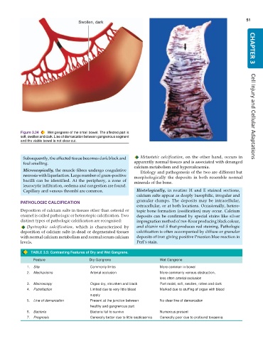
Figure 3.34 Wet gangrene of the small bowel. The affected part is Cell Injury and Cellular Adaptations
soft, swollen and dark. Line of demarcation between gangrenous segment
and the viable bowel is not clear-cut.
Subsequently, the affected tissue becomes dark black and Metastatic calcification, on the other hand, occurs in
foul smelling. apparently normal tissues and is associated with deranged
calcium metabolism and hypercalcaemia.
Microscopically, the muscle fibres undergo coagulative Etiology and pathogenesis of the two are different but
necrosis with liquefaction. Large number of gram-positive morphologically the deposits in both resemble normal
bacilli can be identified. At the periphery, a zone of minerals of the bone.
leucocytic infiltration, oedema and congestion are found.
Capillary and venous thrombi are common. Histologically, in routine H and E stained sections,
calcium salts appear as deeply basophilic, irregular and
granular clumps. The deposits may be intracellular,
PATHOLOGIC CALCIFICATION
extracellular, or at both locations. Occasionally, hetero-
Deposition of calcium salts in tissues other than osteoid or topic bone formation (ossification) may occur. Calcium
enamel is called pathologic or heterotopic calcification. Two deposits can be confirmed by special stains like silver
distinct types of pathologic calcification are recognised: impregnation method of von-Kossa producing black colour,
Dystrophic calcification, which is characterised by and alizarin red S that produces red staining. Pathologic
deposition of calcium salts in dead or degenerated tissues calcification is often accompanied by diffuse or granular
with normal calcium metabolism and normal serum calcium deposits of iron giving positive Prussian blue reaction in
levels. Perl’s stain.
TABLE 3.5: Contrasting Features of Dry and Wet Gangrene.
Feature Dry Gangrene Wet Gangrene
1. Site Commonly limbs More common in bowel
2. Mechanisms Arterial occlusion More commonly venous obstruction,
less often arterial occlusion
3. Macroscopy Organ dry, shrunken and black Part moist, soft, swollen, rotten and dark
4. Putrefaction Limited due to very little blood Marked due to stuffing of organ with blood
supply
5. Line of demarcation Present at the junction between No clear line of demarcation
healthy and gangrenous part
6. Bacteria Bacteria fail to survive Numerous present
7. Prognosis Generally better due to little septicaemia Generally poor due to profound toxaemia

