


Page 72 - Textbook of Pathology, 6th Edition
P. 72
56                                                      hyperplastic growth. Neoplasia differs from hyperplasia in
                                                               having hyperplastic growth with loss of growth-regulatory
                                                               mechanism due to change in genetic composition of the cell.
                                                               Hyperplasia, on the other hand, persists so long as stimulus
                                                               is present.
                                                               CAUSES. As with other non-neoplastic adaptive disorders
                                                               of growth, hyperplasia has also been divided into physiologic
                                                               and pathologic.
     SECTION I
                                                               A. Physiologic hyperplasia. The two most common types
                                                               are as follows:
                                                               1. Hormonal hyperplasia i.e. hyperplasia occurring under the
                                                               influence of hormonal stimulation e.g.
                                                               i) Hyperplasia of female breast at puberty, during preg-
                                                               nancy and lactation.
                                                               ii) Hyperplasia of pregnant uterus.
                                                               iii) Proliferative activity of normal endometrium after a
                                                               normal menstrual cycle.
                                                               iv) Prostatic hyperplasia in old age.
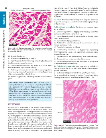
           Figure 3.41  Cardiac hypertrophy. The myocardial muscle fibres are
           thick with abundance of eosinophilic cytoplasm. Nuclei  are also enlarged  2. Compensatory hyperplasia i.e. hyperplasia occurring
           with irregular outlines.                            following removal of part of an organ or a contralateral organ
                                                               in paired organ e.g.
           iii) Intestinal strictures                          i) Regeneration of the liver following partial hepatectomy
           iv) Muscular arteries in hypertension.              ii) Regeneration of epidermis after skin abrasion
           3. Hypertrophy of skeletal muscle e.g. hypertrophied muscles  iii) Following nephrectomy on one side, there is hyperplasia
           in athletes and manual labourers.                   of nephrons of the other kidney.
           4. Compensatory hypertrophy may occur in an organ when  B. Pathologic hyperplasia. Most examples of pathologic
           the contralateral organ is removed e.g.             hyperplasia are due to excessive stimulation of hormones or
           i) Following nephrectomy on one side in a young patient,  growth factors e.g.
     General Pathology and Basic Techniques
           there is compensatory hypertrophy as well as hyperplasia  i) Endometrial hyperplasia following oestrogen excess.
           of the nephrons of the other kidney.                ii) In wound healing, there is formation of granulation tissue
           ii) Adrenal hyperplasia following removal of one adrenal  due to proliferation of fibroblasts and endothelial cells.
           gland.

            MORPHOLOGIC FEATURES. The affected organ is
            enlarged and heavy. For example, a hypertrophied heart
            of a patient with systemic hypertension may weigh
            700-800 g as compared to average normal adult weight of
            350 g. There is enlargement of muscle fibres as well as of
            nuclei (Fig. 3.41). At ultrastructural level, there is increased
            synthesis of DNA and RNA, increased protein synthesis
            and increased number of organelles like mitochondria,
            endoplasmic reticulum and myofibrils.

           HYPERPLASIA
           Hyperplasia is an increase in the number of parenchymal
           cells resulting in enlargement of the organ or tissue. Quite
           often, both hyperplasia and hypertrophy occur together.
           Hyperplasia occurs due to increased recruitment of cells from
           G  (resting) phase of the cell cycle to undergo mitosis, when
            0
           stimulated. All body cells do not possess hyperplastic growth
           potential (Chapter 6). Labile cells (e.g. epithelial cells of the
           skin and mucous membranes, cells of the bone marrow and
           lymph nodes) and stable cells (e.g. parenchymal cells of the
           liver, pancreas, kidney, adrenal, and thyroid) can undergo  Figure 3.42  Pseudocarcinomatous hyperplasia of the skin.  The
           hyperplasia, while permanent cells (e.g. neurons, cardiac and  epidermis shows an increase in the number of layers of the squamous
                                                               epithelium. The intervening dermal soft tissue shows moderate chronic
           skeletal muscle) have little or no capacity for regenerative  inflammation.

