


Page 66 - Textbook of Pathology, 6th Edition
P. 66
50


     SECTION I


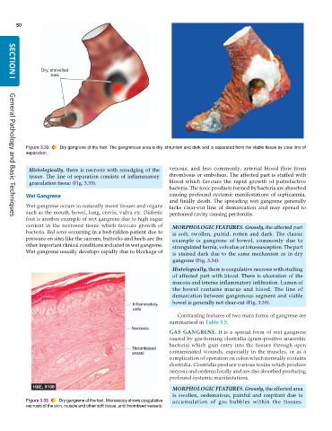
           Figure 3.32  Dry gangrene of the foot. The gangrenous area is dry, shrunken and dark and is separated from the viable tissue by clear line of
           separation.


            Histologically, there is necrosis with smudging of the  venous, and less commonly, arterial blood flow from
            tissue. The line of separation consists of inflammatory  thrombosis or embolism. The affected part is stuffed with
            granulation tissue (Fig. 3.33).                    blood which favours the rapid growth of putrefactive
                                                               bacteria. The toxic products formed by bacteria are absorbed
           Wet Gangrene                                        causing profound systemic manifestations of septicaemia,
                                                               and finally death. The spreading wet gangrene generally
           Wet gangrene occurs in naturally moist tissues and organs  lacks clear-cut line of demarcation and may spread to
           such as the mouth, bowel, lung, cervix, vulva etc. Diabetic  peritoneal cavity causing peritonitis.
           foot is another example of wet gangrene due to high sugar
     General Pathology and Basic Techniques
           content in the necrosed tissue which favours growth of  MORPHOLOGIC FEATURES. Grossly, the affected part
           bacteria. Bed sores occurring in a bed-ridden patient due to  is soft, swollen, putrid, rotten and dark. The classic
           pressure on sites like the sacrum, buttocks and heels are the  example is gangrene of bowel, commonly due to
           other important clinical conditions included in wet gangrene.  strangulated hernia, volvulus or intussusception. The part
           Wet gangrene usually develops rapidly due to blockage of  is stained dark due to the same mechanism as in dry
                                                                 gangrene (Fig. 3.34).
                                                                 Histologically, there is coagulative necrosis with stuffing
                                                                 of affected part with blood. There is ulceration of the
                                                                 mucosa and intense inflammatory infiltration. Lumen of
                                                                 the bowel contains mucus and blood. The line of
                                                                 demarcation between gangrenous segment and viable
                                                                 bowel is generally not clear-cut (Fig. 3.35).

                                                                  Contrasting features of two main forms of gangrene are
                                                               summarised in Table 3.5.

                                                               GAS GANGRENE. It is a special form of wet gangrene
                                                               caused by gas-forming clostridia (gram-positive anaerobic
                                                               bacteria) which gain entry into the tissues through open
                                                               contaminated wounds, especially in the muscles, or as a
                                                               complication of operation on colon which normally contains
                                                               clostridia. Clostridia produce various toxins which produce
                                                               necrosis and oedema locally and are also absorbed producing
                                                               profound systemic manifestations.
                                                                 MORPHOLOGIC FEATURES. Grossly, the affected area
                                                                 is swollen, oedematous, painful and crepitant due to
           Figure 3.33  Dry gangrene of the foot. Microscopy shows coagulative  accumulation of gas bubbles within the tissues.
           necrosis of the skin, muscle and other soft tissue, and thrombsed vessels.

