


Page 69 - Textbook of Pathology, 6th Edition
P. 69
53  CHAPTER 3


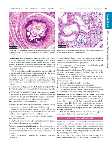
           Figure 3.37  Dystrophic calcification in degenerated tunica media  Figure 3.38  Metastatic calcification in tubular basement membrane  Cell Injury and Cellular Adaptations
           of muscular artery of uterine myometrium in Mönckeberg’s arterio-  in nephrocalcinosis due to hypercalcaemia.
           sclerosis.

           Pathogenesis of dystrophic calcification. It is not quite clear  2. Milk-alkali syndrome caused by excessive oral intake of
           as to how dystrophic calcification takes place. Since serum  calcium in the form of milk and administration of calcium
           calcium levels are within normal limits, the denatured  carbonate in the treatment of peptic ulcer.
           proteins in necrotic or degenerated tissue bind phosphate  3. Hypercalcaemia of infancy is another condition in which
           ions, which react with calcium ions to form precipitates of  metastatic calcification may occur.
           calcium phosphate.                                  Sites of metastatic calcification. Metastatic calcification may
              The process of dystrophic calcification has been likened
           to the formation of normal hydroxyapatite in the bone  occur in any normal tissue of the body but affects the
           involving 2 phases: initiation and propagation.     following organs more commonly:
                                                               1. Kidneys, especially at the basement membrane of tubular
              Initiation is the phase in which precipitates of calcium  epithelium and in the tubular lumina causing nephro-
           phosphate begin to accumulate intracellularly in the  calcinosis (Fig.3.38).
           mitochondria, or extracellularly in membrane-bound  2. Lungs, especially in the alveolar walls.
           vesicles.
                                                               3. Stomach, on the acid-secreting fundal glands.
              Propagation is the phase in which minerals deposited in  4. Blood vessels, especially on the internal elastic lamina.
           the initiation phase are propagated to form mineral crystals.  5. Cornea is another site affected by metastatic calcification.
           METASTATIC CALCIFICATION.  Since metastatic calcifi-  6. Synovium of the joint causing pain and dysfunction.
           cation occurs in normal tissues due to hypercalcaemia, its  Pathogenesis  of  metastatic calcification.  Metasatic
           causes would include one of the following two conditions:  calcification at the above-mentioned sites occurs due to
              Excessive mobilisation of calcium from the bone.  excessive binding of inorganic phosphate ions with calcium
              Excessive absorption of calcium from the gut.    ions, which are elevated due to underlying metabolic
                                                               derangement. This leads to formation of precipitates of
           Excessive mobilisation of calcium from the bone. These  calcium phosphate at the preferential sites. Metastatic
           causes are more common and include the following:   calcification is reversible upon correction of underlying
           1. Hyperparathyroidism which may be primary such as due  metabolic disorder.
           to parathyroid adenoma, or secondary such as from      The distinguishing features between the two types of
           parathyroid hyperplasia, chronic renal failure etc.  pathologic calcification are summarised in Table 3.6.
           2. Bony destructive  lesions such as multiple myeloma,
           metastatic carcinoma.                                          CELLULAR ADAPTATIONS
           3. Prolonged immobilisation of a patient results in disuse  For the sake of survival on exposure to stress, the cells make
           atrophy of the bones and hypercalcaemia.            adjustments with the changes in their environment (i.e.
           Excessive absorption of calcium from the gut. Less often,  adapt) to the physiologic needs (physiologic adaptation) and
           excess calcium may be absorbed from the gut causing  to non-lethal pathologic injury (pathologic adaptation). Broadly
           hypercalcaemia and metastatic calcification. These causes are  speaking, such physiologic and pathologic adaptations occur
           as under:                                           by following processes (Fig. 3.39):
           1. Hypervitaminosis D results in increased calcium     Decreasing or increasing their size i.e.  atrophy and
           absorption.                                         hypertrophy respectively, or by increasing their number i.e.

