
Page 748 - Textbook of Pathology, 6th Edition
P. 748
732 5. At perimenopause: endometrial hyperplasia, carcinoma,
polyps and senile atrophy.
It has been observed that women who ovulate may also
occasionally have anovulatory cycles. In addition to
anovulatory cycles, DUB may occur in inadequate luteal phase
that manifests clinically as infertility (ovulatory dysfunctional
bleeding). In such cases, the premenstrual endometrial biopsy
shows histologic lag of more than 2 days.
ENDOMETRITIS AND MYOMETRITIS
Inflammatory involvement of the endometrium and
myometrium are uncommon clinical problems; myometritis
is seen less frequently than endometritis and occurs in
continuation with endometrial infections. Endometritis and
myometritis may be acute or chronic.
Acute form generally results from 3 types of causes—
puerperal (following full-term delivery, abortion and
retained products of conception), intrauterine contraceptive
device (IUCD), and extension of gonorrheal infection from
the cervix and vagina.
Chronic form is more common and occurs by the same
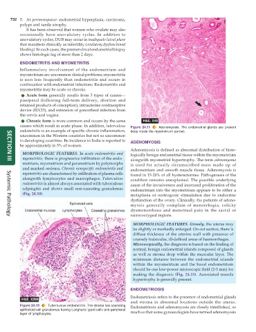
causes which result in acute phase. In addition, tuberculous Figure 24.11 Adenomyosis. The endometrial glands are present
endometritis is an example of specific chronic inflammation, deep inside the myometrium (arrow).
uncommon in the Western countries but not so uncommon
in developing countries. Its incidence in India is reported to ADENOMYOSIS
be approximately in 5% of women.
Adenomyosis is defined as abnormal distribution of histo-
MORPHOLOGIC FEATURES. In acute endometritis and logically benign endometrial tissue within the myometrium
myometritis, there is progressive infiltration of the endo- alongwith myometrial hypertrophy. The term adenomyoma
metrium, myometrium and parametrium by polymorphs is used for actually circumscribed mass made up of
SECTION III
and marked oedema. Chronic nonspecific endometritis and endometrium and smooth muscle tissue. Adenomyosis is
myometritis are characterised by infiltration of plasma cells found in 15-20% of all hysterectomies. Pathogenesis of the
alongwith lymphocytes and macrophages. Tuberculous condition remains unexplained. The possible underlying
endometritis is almost always associated with tuberculous cause of the invasiveness and increased proliferation of the
salpingitis and shows small non-caseating granulomas endometrium into the myometrium appears to be either a
(Fig. 24.10). metaplasia or oestrogenic stimulation due to endocrine
dysfunction of the ovary. Clinically, the patients of adeno-
myosis generally complain of menorrhagia, colicky
dysmenorrhoea and menstrual pain in the sacral or
sacrococcygeal regions.
Systemic Pathology
MORPHOLOGIC FEATURES. Grossly, the uterus may
be slightly or markedly enlarged. On cut section, there is
diffuse thickness of the uterine wall with presence of
coarsely trabecular, ill-defined areas of haemorrhages.
Microscopically, the diagnosis is based on the finding of
normal, benign endometrial islands composed of glands
as well as stroma deep within the muscular layer. The
minimum distance between the endometrial islands
within the myometrium and the basal endometrium
should be one low-power microscopic field (2-3 mm) for
making the diagnosis (Fig. 24.11). Associated muscle
hypertrophy is generally present.
ENDOMETRIOSIS
Endometriosis refers to the presence of endometrial glands
and stroma in abnormal locations outside the uterus.
Figure 24.10 Tuberculous endometritis. The stroma has caseating Endometriosis and adenomyosis are closely interlinked, so
epithelioid cell granulomas having Langhans’ giant cells and peripheral much so that some gynaecologists have termed adenomyosis
layer of lymphocytes.