
Page 749 - Textbook of Pathology, 6th Edition
P. 749
as endometriosis interna and the other category termed as common site of endometriosis and shows numerous cysts 733
endometriosis externa for similar appearance at the extrauterine varying in diameter from 0.1 to 2.5 cm. Ovarian
sites. However, the two differ as regards age, fertility and involvement is often bilateral. Larger cysts, 3-5 cm in
histogenesis and thus endometriosis should be regarded as diameter, filled with old dark brown blood form ‘chocolate
a distinct clinicopathologic entity. cysts’ of the ovary.
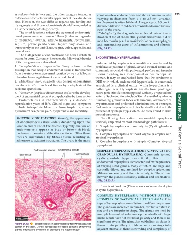
The chief locations where the abnormal endometrial Histologically, the diagnosis is simple and rests on ident-
development may occur are as follows (in descending order ification of foci of endometrial glands and stroma, old or
of frequency): ovaries, uterine ligaments, rectovaginal new haemorrhages, haemosiderin-laden macrophages
septum, pelvic peritoneum, laparotomy scars, and and surrounding zone of inflammation and fibrosis
infrequently in the umbilicus, vagina, vulva, appendix and (Fig. 24.12).
hernial sacs.
The histogenesis of endometriosis has been a debatable
matter for years. Currently, however, the following 3 theories ENDOMETRIAL HYPERPLASIAS
of its histogenesis are described: Endometrial hyperplasia is a condition characterised by
1. Transplantation or regurgitation theory is based on the proliferative patterns of glandular and stromal tissues and
assumption that ectopic endometrial tissue is transplanted commonly associated with prolonged, profuse and irregular
from the uterus to an abnormal location by way of fallopian uterine bleeding in a menopausal or postmenopausal
tubes due to regurgitation of menstrual blood. woman. It may be emphasised here that the syndrome of
2. Metaplastic theory suggests that ectopic endometrium DUB with which endometrial hyperplasia is commonly
develops in situ from local tissues by metaplasia of the associated is a clinical entity, while hyperplasia is a
coelomic epithelium. pathologic term. Hyperplasia results from prolonged
3. Vascular or lymphatic dissemination explains the develop- oestrogenic stimulation unopposed with any progestational
ment of endometrial tissue at extrapelvic sites by these routes. activity. Such conditions include Stein-Leventhal syndrome,
Endometriosis is characteristically a disease of functioning granulosa-theca cell tumours, adrenocortical
reproductive years of life. Clinical signs and symptoms hyperfunction and prolonged administration of oestrogen.
include intrapelvic bleeding from implants, severe Endometrial hyperplasia is clinically significant due to the CHAPTER 24
dysmenorrhoea, pelvic pain, dyspareunia and infertility. presence of cytologic atypia which is closely linked to endo-
metrial carcinoma.
MORPHOLOGIC FEATURES. Grossly, the appearance The following classification of endometrial hyperplasias
of endometriosis varies widely depending upon the is widely employed by most gynaecologic pathologists:
location and extent of the disease. Typically, the foci of 1. Simple hyperplasia without atypia (Cystic glandular
endometriosis appear as blue or brownish-black hyperplasia).
underneath the surface of the sites mentioned. Often, these 2. Complex hyperplasia without atypia (Complex non-
foci are surrounded by fibrous tissue resulting in atypical hyperplasia).
adherence to adjacent structures. The ovary is the most 3. Complex hyperplasia with atypia (Complex atypical
hyperplasia). The Female Genital Tract
SIMPLE HYPERPLASIA WITHOUT ATYPIA (CYSTIC
GLANDULAR HYPERPLASIA). Commonly termed
cystic glandular hyperplasia (CGH), this form of
endometrial hyperplasia is characterised by the presence
of varying-sized glands, many of which are large and
cystically dilated and are lined by atrophic epithelium.
Mitoses are scanty and there is no atypia. The stroma
between the glands is sparsely cellular and oedematous
(Fig. 24.13,A).
There is minimal risk (1%) of adenocarcinoma developing
in cystic hyperplasia.
COMPLEX HYPERPLASIA WITHOUT ATYPIA
(COMPLEX NON-ATYPICAL HYPERPLASIA). This
type of hyperplasia shows distinct proliferative pattern.
The glands are increased in number, exhibit variation in
size and are irregular in shape. The glands are lined by
multiple layers of tall columnar epithelial cells with large
nuclei which have not lost basal polarity and there is no
significant atypia. The glandular epithelium at places is
Figure 24.12 Endometriosis in abdominal scar following caesarean thrown into papillary infolds or out-pouchings into
section in the past. Dense fibrocollagenic tissue contains endometrial
glands, stroma and evidence of preceding old haemorrhage. adjacent stroma i.e. there is crowding and complexity of