
Page 753 - Textbook of Pathology, 6th Edition
P. 753
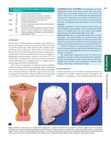
TABLE 24.5: FIGO Clinical Staging of Carcinoma of the MORPHOLOGIC FEATURES. Leiomyomas are most 737
Endometrium. frequently located in the uterus where they may occur
Stage IA Tumour limited to endometrium. within the myometrium (intramural or interstitial), the
IB Invasion to less than one-half the myometrium. serosa (subserosal), or just underneath the endometrium
IC Invasion to more than one-half the myometrium. (submucosal). Subserosal and submucosal leiomyomas
Stage IIA Endocervical glandular involvement only. may develop pedicles and protrude as pedunculated
IIB Cervical stromal invasion. myomas. Leiomyomas may involve the cervix or broad
Stage IIIA Tumour invades serosa and/or adnexa and/or positive ligament.
peritoneal cytology. Grossly, irrespective of their location, leiomyomas are
IIIB Metastases to pelvic and/or para-aortic lymph nodes. often multiple, circumscribed, firm, nodular, grey-white
Stage IVA Tumour invasion of bladder and/or bowel mucosa. masses of variable size. On cut section, they exhibit
IVB Distant metastases including intra-abdominal and/or characteristic whorled pattern (Fig. 24.16, A,B).
inguinal lymph nodes.
Histologically, they are essentially composed of 2 tissue
elements—whorled bundles of smooth muscle cells
Leiomyoma admixed with variable amount of connective tissue. The
smooth muscle cells are uniform in size and shape with
Leiomyomas or fibromyomas, commonly called fibroids by abundant cytoplasm and central oval nuclei (Fig. 24.17).
the gynaecologists, are the most common uterine tumours Cellular leiomyoma has preponderance of smooth
of smooth muscle origin, often admixed with variable amount muscle elements and may superficially resemble leiomyo-
of fibrous tissue component. About 20% of women above sarcoma but is distinguished from it by the absence of
the age of 30 years harbour uterine myomas of varying size. mitoses (see below).
Vast majority of them are benign and cause no symptoms. The pathologic appearance may be altered by
Malignant transformation occurs in less than 0.5% of secondary changes in the leiomyomas; these include:
leiomyomas. Symptomatic cases may produce abnormal hyaline degeneration, cystic degeneration, infarction,
uterine bleeding, pain, symptoms due to compression of calcification, infection and suppuration, necrosis, fatty
surrounding structures and infertility. change, and rarely, sarcomatous change. CHAPTER 24
The cause of leiomyomas is unknown but the possible
stimulus to their proliferation is oestrogen. This is evidenced
by increase in their size in pregnancy (Fig. 24.16,C) and high Leiomyosarcoma
dose oestrogen-therapy and their regression following Leiomyosarcoma is an uncommon malignant tumour as
menopause and castration. Other possible factors implicated compared to its rather common benign counterpart. The
in its etiology are human growth hormone and sterility. incidence of malignancy in pre-existing leiomyoma is less The Female Genital Tract
Figure 24.16 Leiomyomas. A, Diagrammatic appearance of common locations and characteristic whorled appearance on cut section.
B, Sectioned surface of the uterus shows multiple circumscribed, firm nodular masses of variable sizes—submucosal (white arrows) and intramural
(black arrows) in location having characteristic whorling. C, The opened up uterine cavity shows an intrauterine gestation sac with placenta (white
arrow) and a single circumscribed, enlarged, firm nodular mass in intramural location (black arrow) having grey-white whorled pattern.