
Page 754 - Textbook of Pathology, 6th Edition
P. 754
738
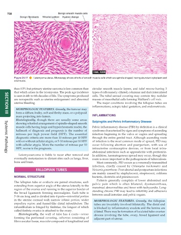
Figure 24.17 Leiomyoma uterus. Microscopy shows whorls of smooth muscle cells which are spindle-shaped, having abundant cytoplasm and
oval nuclei.
than 0.5% but primary uterine sarcoma is less common than circular smooth muscle layers, and tubal mucosa having 3
that which arises in the leiomyoma. The peak age incidence types of cells namely: ciliated, columnar and dark intercalated
is seen in 4th to 6th decades of life. The symptoms produced cells. The tubal serosal covering may contain tiny nodular
are nonspecific such as uterine enlargement and abnormal masses of mesothelial cells forming Walthard’s cell rests.
uterine bleeding. The major conditions involving the fallopian tubes are
inflammations, ectopic tubal gestation, and endometriosis.
MORPHOLOGIC FEATURES. Grossly, the tumour may
form a diffuse, bulky, soft and fleshy mass, or a polypoid
SECTION III
mass projecting into lumen. INFLAMMATIONS
Histologically, though there are usually some areas Salpingitis and Pelvic Inflammatory Disease
showing whorled arrangement of spindle-shaped smooth
muscle cells having large and hyperchromatic nuclei, the Pelvic inflammatory disease (PID) by definition is a clinical
hallmark of diagnosis and prognosis is the number of syndrome characterised by signs and symptoms of ascending
mitoses per high power field (HPF). The essential infection beginning in the vulva or vagina and spreading
diagnostic criteria are: more than 10 mitoses per 10 HPF through the entire genital tract. Although ascending route
with or without cellular atypia, or 5-10 mitoses per 10 HPF of infection is the most common mode of spread, PID may
with cellular atypia. More the number of mitoses per 10 occur following abortion and puerperium, with use of
HPF, worse is the prognosis. intrauterine contraceptive devices, or from local intra-
abdominal infections such as appendicitis with peritonitis.
Leiomyosarcoma is liable to recur after removal and In addition, haematogenous spread may occur, though this
Systemic Pathology
eventually metastasises to distant sites such as lungs, liver, route is more important in the pathogenesis of tuberculosis.
bone and brain. Most commonly, PID occurs as a venereally-transmitted
infection, chiefly caused by Chlamydia trachomatis and
FALLOPIAN TUBES Neisseria gonorrhoeae. Post-abortal and postpartum infections
are mainly caused by staphylococci, streptococci, coliform
NORMAL STRUCTURE bacteria, clostridia and pneumococci.
Patients generally complain of lower abdominal and
The fallopian tube or oviducts are paired structures, each pelvic pain which is often bilateral, dysmenorrhoea,
extending from superior angle of the uterus laterally to the menstrual abnormalities and fever with tachycardia. Long-
region of the ovaries and running in the superior border of standing chronic PID may lead to infertility and adhesions
the broad ligaments forming mesosalpinx. Each tube is between small intestine and pelvic organs.
7-14 cm long and is divided into 4 parts—interstitial portion
in the uterine cornual wall; narrow isthmic portion; wider MORPHOLOGIC FEATURES. Grossly, the fallopian
ampullary region; and funnel-like distal infundibulum. The tubes are invariably involved bilaterally. The distal end
infundibulum is fringed by fimbriae, the longest of which is blocked by inflammatory exudate and the lumina are
called fimbria ovarica is attached to the ovary. dilated. There may be formation of loculated tubo-ovarian
Histologically, the wall of tube has 4 coats—serous abscess involving the tube, ovary, broad ligament and
forming the peritoneal covering, subserous consisting of adjacent part of uterus.
fibrovascular tissue, muscular composed of longitudinal and