
Page 756 - Textbook of Pathology, 6th Edition
P. 756
740
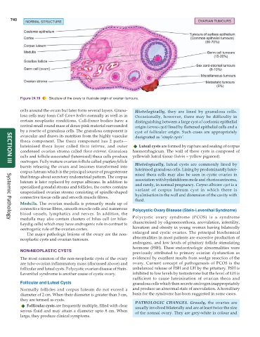
Figure 24.19 Structure of the ovary to illustrate origin of ovarian tumours.
cells around the ovum but later form several layers. Granu- Histologically, they are lined by granulosa cells.
losa cells may form Call-Exner bodies normally as well as in Occasionally, however, there may be difficulty in
certain neoplastic conditions. Call-Exner bodies have a distinguishing between a large cyst of coelomic epithelial
central small round mass of dense pink material surrounded origin (serous cyst) lined by flattened epithelial cells and a
by a rosette of granulosa cells. The granulosa component is cyst of follicular origin. Such cases are appropriately
avascular and draws its nutrition from the highly vascular designated as ‘simple cysts’.
theca component. The theca component has 2 parts—
luteinised theca layer called theca interna, and outer Luteal cysts are formed by rupture and sealing of corpus
condensed ovarian stroma called theca externa. Granulosa haemorrhagicum. The wall of these cysts is composed of
cells and follicle-associated (luteinised) theca cells produce yellowish luteal tissue (lutein = yellow pigment).
oestrogen. Fully mature ovarian follicle called graafian follicle Histologically, luteal cysts are commonly lined by
SECTION III
bursts releasing the ovum and becomes transformed into luteinised granulosa cells. Lining by predominantly lutei-
corpus luteum which is the principal source of progesterone nised theca cells may also be seen in cystic ovaries in
that brings about secretory endometrial pattern. The corpus association with hydatidiform mole and choriocarcinoma,
luteum is later replaced by corpus albicans. In addition to and rarely, in normal pregnancy. Corpus albicans cyst is a
specialised gonadal stroma and follicles, the cortex contains variant of corpus luteum cyst in which there is
unspecialised ovarian stroma consisting of spindle-shaped hyalinisation in the wall and distension of the cavity with
connective tissue cells and smooth muscle fibres. fluid.
Medulla. The ovarian medulla is primarily made up of
connective tissue fibres, smooth muscle cells and numerous Polycystic Ovary Disease (Stein-Leventhal Syndrome)
blood vessels, lymphatics and nerves. In addition, the
medulla may also contain clusters of hilus cell (or hilar- Polycystic ovary syndrome (PCOS) is a syndrome
Systemic Pathology
Leydig cells) which may have androgenic role in contrast to characterised by oligomenorrhoea, anovulation, infertility,
oestrogenic role of the ovarian cortex. hirsutism and obesity in young women having bilaterally
The major pathologic lesions of the ovary are the non- enlarged and cystic ovaries. The principal biochemical
neoplastic cysts and ovarian tumours. abnormalities in most patients are excessive production of
androgens, and low levels of pituitary follicle stimulating
hormone (FSH). These endocrinologic abnormalities were
NON-NEOPLASTIC CYSTS previously attributed to primary ovarian dysfunction as
The most common of the non-neoplastic cysts of the ovary evidenced by excellent results from wedge resection of the
are tubo-ovarian inflammatory mass (discussed above) and ovary. Current concept of pathogenesis of PCOS is the
follicular and luteal cysts. Polycystic ovarian disease of Stein- unbalanced release of FSH and LH by the pituitary. FSH is
Leventhal syndrome is another cause of cystic ovary. inhibited to low levels by testosterone but the level of LH is
sufficient to cause luteinisation of ovarian theca and
Follicular and Luteal Cysts granulosa cells which then secrete androgen inappropriately
Normally follicles and corpus luteum do not exceed a and produce an abnormal state of anovulation. A hereditary
diameter of 2 cm. When their diameter is greater than 3 cm, basis for the syndrome has been suggested in some cases.
they are termed as cysts. PATHOLOGIC CHANGES. Grossly, the ovaries are
Follicular cysts are frequently multiple, filled with clear usually involved bilaterally and are at least twice the size
serous fluid and may attain a diameter upto 8 cm. When of the normal ovary. They are grey-white in colour and
large, they produce clinical symptoms.