
Page 755 - Textbook of Pathology, 6th Edition
P. 755
Microscopically, the appearance varies with the duration 739
of inflammatory process.
The process begins with acute salpingitis characterised
by oedema and intense acute inflammatory infiltrate of
neutrophils involving the tubal mucosa as well as wall.
The lumen is filled with purulent exudate consisting of
leucocytes and sloughed off epithelial cells.
The purulent process may extend to involve tube as
well as ovary causing salpingo-oophoritis and forming
tubo-ovarian abscess.
The escape of purulent exudate into the peritoneal
cavity produces pelvic peritonitis and pelvic abscess.
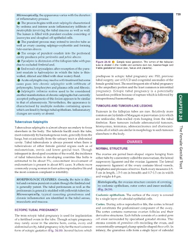
Pyosalpinx is distension of the fallopian tube with pus Figure 24.18 Ectopic tubal gestation. The lumen of the fallopian
due to occluded fimbrial end. tube is dilated in the middle and contains dark tan, haemorrhagic and
End-result of pyosalpinx after resorption of the puru- friable gestational tissue (sac, foetus and placenta).
lent exudate is hydrosalpinx in which the tube is thin-
walled, dilated and filled with clear watery fluid. predispose to ectopic tubal pregnancy are: PID, previous
Acute salpingitis may resolve with treatment but some tubal surgery, use of IUCD and congenital anomalies of the
cases pass into chronic salpingitis with infiltrate of female genital tract. The most frequent site of tubal pregnancy
polymorphs, lymphocytes and plasma cells and fibrosis. is the ampullary portion and the least common is interstitial
Salpingitis isthmica nodosa used to be considered pregnancy. Ectopic tubal pregnancy is a potentially
another manifestation of chronic salpingitis but currently hazardous problem because of rupture which is followed by
accepted pathogenesis of this lesion appears to be similar intraperitoneal haemorrhage.
to that of adenomyosis. Nevertheless, the appearance is
characterised by multiple nodules containing spaces TUMOURS AND TUMOUR-LIKE LESIONS
which are lined by benign tubal epithelium. Inflammatory Tumours in the fallopian tubes are rare. Relatively more CHAPTER 24
changes are scanty or absent.
common are hydatids of Morgagni or parovarian cysts which
are unilocular, thin-walled cysts hanging from the tubal
Tuberculous Salpingitis fimbriae. Rare tumours include adenomatoid tumours,
Tuberculous salpingitis is almost always secondary to focus leiomyomas, teratomas, adenocarcinomas and choriocarci-
elsewhere in the body. The tubercle bacilli reach the tube, noma all of which are similar in morphology to such tumours
most commonly by haematogenous route, generally from the elsewhere in the body.
lungs, but occasionally from the urinary tract or abdominal
cavity. Tubal tuberculosis is always present when there is OVARIES
tuberculosis of other female genital organs such as of
endometrium, cervix and lower genital tract. Though NORMAL STRUCTURE The Female Genital Tract
infrequent in developed countries of the world, the incidence The ovaries are paired bean-shaped organs hanging from
of tubal tuberculosis in developing countries like India is either tube by a mesentery called the mesovarium, the lateral
estimated to be about 5%; concomitant involvement of suspensory ligament and the ovarian ligament. The lateral
endometrium is present in about 80% cases. It affects more suspensory ligament of the ovary contains blood vessels,
commonly young women in their active reproductive life and lymphatics and plexuses of nerves. Each ovary measures 2.5-
the most common complaint is infertility. 5 cm in length, 1.5-3 cm in breadth and 0.7-1.5 cm in width
and weighs 4-8 gm.
MORPHOLOGIC FEATURES. Grossly, the tube is dila-
ted and contains purulent exudate though the fimbrial end Histologically, the ovarian structure consists of covering
is generally patent. The tubal peritoneum as well as the by coelomic epithelium, outer cortex and inner medulla
peritoneum in general is studded with yellowish tubercles. (Fig. 24.19).
Microscopically, typical caseating granulomas and Coelomic epithelium. The surface of the ovary is covered
chronic inflammation are identified in the tubal serosa, by a single layer of cuboidal epithelial cells.
muscularis and mucosa.
Cortex. During active reproductive life, the cortex is broad
and constitutes the predominant component of the ovary.
ECTOPIC TUBAL PREGNANCY
The cortex contains numerous ovarian follicles and their
The term ectopic tubal pregnancy is used for implantation derivative structures. Each follicle consists of a central germ
of a fertilised ovum in the tube. Though ectopic pregnancy cell ovum surrounded by specialised gonadal stroma. This
may rarely occur in the uterine horn, cornu, ovary and stroma consists of granulosa cells encircling the ovum, and
abdominal cavity, tubal pregnancy is by far the most common concentrically-arranged plump spindle-shaped theca cells. In
form of ectopic gestation (Fig. 24.18). Several factors which infancy, the granulosa cells form a single layer of cuboidal