
Page 797 - Textbook of Pathology, 6th Edition
P. 797
781
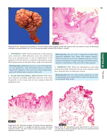
Figure 26.18 Squamous cell papilloma. A, The skin surface shows a papillary growth with a pedicle while the surface is smooth. B, Microscopy
resembles verruca but differs from it by not having vacuolated koilocytic cells in stratum malpighii.
1. EPIDERMAL CYST. These intradermal or subcutaneous Histologically, the cyst wall is composed of palisading
cysts, commonly called sebaceous cysts, are common and squamous epithelial cells. These cells undergo degene-
may occur spontaneously or due to implantation of the ration towards the cyst cavity. Rupture of the cyst wall is
epidermis into the dermis or subcutis (implantation cysts). common and leads to foreign body giant cell inflammatory CHAPTER 26
Most frequent sites are the skin of face, scalp, neck and trunk. reaction. Calcification in the cyst wall is often present.
Histologically, epidermal cysts have a cyst wall composed
of true epidermis with laminated layers of keratin 3. DERMOID CYST. These are subcutaneous cysts often
(Fig. 26.21). Rupture of the cyst may incite foreign body present since birth. Dermoid cysts are more common on the
giant cell inflammatory reaction in the wall. face, along the lines of embryonic closure
2. PILAR (TRICHILEMMAL, SEBACEOUS) CYST. These Histologically, the cyst wall contains epidermis as well The Skin
cysts clinically resemble epidermal cysts but occur more as appendages such as hair follicles, sebaceous glands and
frequently on the scalp and are less common than the sweat glands (Fig. 26.22).
epidermal cysts.
Figure 26.19 Seborrheic keratosis. The border of the elevated lesion
at the lateral margin is in a straight line from the normal uninvolved Figure 26.20 Fibroepithelial polyp (acrochordon or soft fibroma).
epidermis (arrow). The other features include papillomatosis, hyper- The epidermis is raised as polypoid mass over dense hyalinised fibrous
keratosis and acanthosis. connective tissue in the dermis.