
Page 828 - Textbook of Pathology, 6th Edition
P. 828
812 ETIOPATHOGENESIS. Most important risk factor translocation between 2 genes—PAX-8 (paired domain
implicated in the etiology of thyroid cancer is external transcription factor) and PPARγ-1 (gene coding for
radiation, and to a some extent there is role of TSH receptors peroxisome proliferator-activator receptor γ-1), has also been
and iodine excess, while pathogenesis of thyroid cancer is described in a proportion of cases of follicular thyroid
explained on genetic alterations. neoplasms, both adenoma and carcinoma.
1. External radiation. The single most important iii) Medullary thyroid carcinoma: Medullary thyroid carcinoma
environmental factor associated with increased risk of arises from parafollicular C-cells in the thyroid. Point
developing thyroid carcinoma after many years of exposure mutation in RET-protooncogene is seen in both familial as a
to external radiation of high dose. Evidences in support well as sporadiac cases of medullary thyroid carcinoma.
include: high incidence of thyroid cancer in individuals iv) Anaplastic thyroid carcinoma: This tumour either arises from
irradiated in early age for enlarged thymus and for skin further dedifferentiation of differentiated papillary or
disorders, in Japanese atomic bomb survivors, and in follicular thyroid carcinoma, or by inactivating point
individuals living in the vicinity of nuclear accident sites. In mutation in p53 tumour suppressor gene or by mutation in
particular, exposure to radiation to children and young adults gene coding for β-catenin pathway.
has been found to be associated with higher incidence of Papillary Thyroid Carcinoma
development of papillary carcinoma later.
2. Iodine excess and TSH. In regions where endemic goitre Papillary carcinoma is the most common type of thyroid
carcinoma, comprising 75-85% of cases. It can occur at all
is widespread, addition of iodine to diet has resulted in ages including children and young adults but the incidence
increase in incidence of papillary cancer. Many well- is higher with advancing age. The tumour is found about
differentiated thyroid cancers express TSH receptors and thus three times more frequently in females than in males.
respond to T suppression of TSH. Papillary carcinoma is typically a slow-growing
4
3. Genetic basis. Familial clustering of thyroid cancer has malignant tumour, most often presenting as an asymptomatic
been observed, especially in medullary carcinoma. Molecular solitary nodule. Involvement of the regional lymph nodes is
studies reveal that thyroid carcinoma is a multistep process common but distant metastases to organs are rare. Some cases
involving genetic alterations but distinct mutations are seen first come to attention by spread to regional lymph nodes
in different histologic types: and cause cervical lymphadenopathy. ‘Lateral aberrant thyroid’
i) Papillary thyroid carcinoma: Mutation in RET gene (gene is the term used for occurrence of thyroid tissue in the lateral
overexpression) located on chromosome 10q is seen in about cervical lymph node, which in most patients represents a
20% cases of papillary thyroid carcinoma. This mutation well-differentiated metastasis of an occult papillary
SECTION III
renders the tyrosine kinase receptor under the target of other carcinoma of the thyroid.
tumour-promoting factors such as radiation exposure in MORPHOLOGIC FEATURES. Grossly, papillary carci-
papillary carcinoma. Another genetic abnormality seen in noma may range from microscopic foci to nodules upto
5-10% cases of papillary thyroid carcinoma is gene 10 cm in diameter and is generally poorly delineated. Cut
rearrangement in NTRK1 (neurotrophic tyrosine kinase surface of the tumour is greyish-white, hard and scar-like
receptor 1 located on chromosome 1q) gene. (Fig. 27.16). Sometimes the tumour is transformed into a
ii) Follicular thyroid carcinoma: About 50% cases of follicular cyst, into which numerous papillae project and is termed
thyroid carcinoma have mutation in RAS family of oncogenes
that includes HRAS, NRAS and KRAS. Besides, fusion- papillary cystadenocarcinoma.
Systemic Pathology
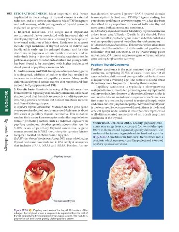
Figure 27.16 Papillary carcinoma of the thyroid. Cut surface of the
enlarged thyroid gland shows a single nodule separated from the rest of
thyroid parenchyma by incomplete fibrous septa (arrow). The nodule is
grey-white soft and shows grossly visible papillary pattern.