
Page 829 - Textbook of Pathology, 6th Edition
P. 829
813
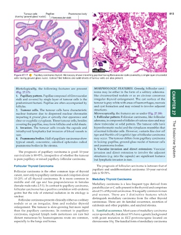
Figure 27.17 Papillary carcinoma thyroid. Microscopy shows branching papillae having flbrovascular stalk covered by a single layer of cuboidal
cells having ground-glass nuclei. Colloid-filled follicles and solid sheets of tumour cells are also present.
Histologically, the following features are present MORPHOLOGIC FEATURES. Grossly, follicular carci-
(Fig. 27.17): noma may be either in the form of a solitary adenoma-
1. Papillary pattern. Papillae composed of fibrovascular like circumscribed nodule or as an obvious cancerous
stalk and covered by single layer of tumour cells is the irregular thyroid enlargement. The cut surface of the
predominant feature. Papillae are often accompanied by tumour is grey-white with areas of haemorrhages, necrosis CHAPTER 27
follicles. and cyst formation and may extend to involve adjacent
2. Tumour cells. The tumour cells have characteristic structures.
nuclear features due to dispersed nuclear chromatin Microscopically, the features are as under (Fig. 27.18):
imparting it ground glass or optically clear appearance and 1. Follicular pattern: Follicular carcinoma, like follicular
clear or oxyphilic cytoplasm. These tumour cells, besides adenoma, is composed of follicles of various sizes and may
covering the papillae, may form follicles and solid sheets. show trabecular or solid pattern. The tumour cells have
3. Invasion. The tumour cells invade the capsule and hyperchromatic nuclei and the cytoplasm resembles that
intrathyroid lymphatics but invasion of blood vessels is of normal follicular cells. However, variants like clear cell
rare. type and Hurthle cell (oxyphilic) type of follicular carcinoma
4. Psammoma bodies. Half of papillary carcinomas show may occur. The tumour differs from papillary carcinoma The Endocrine System
typical small, concentric, calcified spherules called in lacking: papillae, ground-glass nuclei of tumour cells
psammoma bodies in the stroma. and psammoma bodies.
2. Vascular invasion and direct extension: Vascular
The prognosis of papillary carcinoma is good: 10-year invasion and direct extension to involve the adjacent
survival rate is 80-95%, irrespective of whether the tumour structures (e.g. into the capsule) are significant features
is pure papillary or mixed papillary-follicular carcinoma. but lymphatic invasion is rare.
Follicular Thyroid Carcinoma The prognosis of follicular carcinoma is between that of
papillary and undifferentiated carcinoma: 10-year survival
Follicular carcinoma is the other common type of thyroid rate is 50-70%.
cancer, next only to papillary carcinoma and comprises about
10-20% of all thyroid carcinomas. It is more common in Medullary Thyroid Carcinoma
middle and old age and has preponderance in females Medullary carcinoma is a less frequent type derived from
(female-male ratio 2.5:1). In contrast to papillary carcinoma, parafollicular or C-cells present in the thyroid and comprises
follicular carcinoma has a positive correlation with endemic about 5% of thyroid carcinomas. It is equally common in men
goitre but the role of external radiation in its etiology is and women. There are 3 distinctive features which
unclear. distinguish medullary carcinoma from the other thyroid
Follicular carcinoma presents clinically either as a solitary
nodule or as an irregular, firm and nodular thyroid carcinomas. These are: its familial occurrence, secretion of
calcitonin and other peptides, and amyloid stroma.
enlargement. The tumour is slow-growing but more rapid
than the papillary carcinoma. In contrast to papillary 1. Familial occurrence. Most cases of medullary carcinoma
carcinoma, regional lymph node metastases are rare but occur sporadically, but about 10% have a genetic background
distant metastases by haematogenous route are common, with point mutation in RET-protooncogene located on
especially to the lungs and bones. chromosome 10q. The familial form of medullary carcinoma