
Page 862 - Textbook of Pathology, 6th Edition
P. 862
846 (Fig. 28.16). However, sometimes distinction between a
well-differentiated chondrosarcoma and a benign
chondroma may be difficult and in such cases location,
clinical features and radiological appearance are often
helpful.
Rare variants of chondrosarcoma are mesenchymal
chondrosarcoma, dedifferentiated chondrosarcoma and clear
cell chondrosarcoma.
GIANT CELL TUMOUR (OSTEOCLASTOMA)
Giant cell tumour or osteoclastoma is a distinctive neoplasm
with uncertain histogenesis and hence classified separately.
The tumour arises in the epiphysis of long bones close to the
articular cartilage. Most common sites of involvement are
lower end of femur and upper end of tibia (i.e. about the
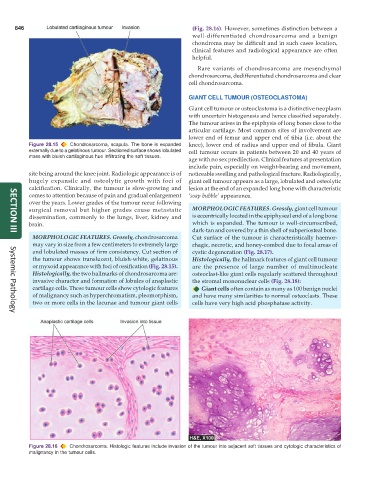
Figure 28.15 Chondrosarcoma, scapula. The bone is expanded knee), lower end of radius and upper end of fibula. Giant
externally due to a gelatinous tumour. Sectioned surface shows lobulated cell tumour occurs in patients between 20 and 40 years of
mass with bluish cartilaginous hue infiltrating the soft tissues.
age with no sex predilection. Clinical features at presentation
include pain, especially on weight-bearing and movement,
site being around the knee joint. Radiologic appearance is of noticeable swelling and pathological fracture. Radiologically,
hugely expansile and osteolytic growth with foci of giant cell tumour appears as a large, lobulated and osteolytic
calcification. Clinically, the tumour is slow-growing and lesion at the end of an expanded long bone with characteristic
comes to attention because of pain and gradual enlargement ‘soap bubble’ appearance.
over the years. Lower grades of the tumour recur following
surgical removal but higher grades cause metastatic MORPHOLOGIC FEATURES. Grossly, giant cell tumour
dissemination, commonly to the lungs, liver, kidney and is eccentrically located in the epiphyseal end of a long bone
brain. which is expanded. The tumour is well-circumscribed,
dark-tan and covered by a thin shell of subperiosteal bone.
SECTION III
MORPHOLOGIC FEATURES. Grossly, chondrosarcoma Cut surface of the tumour is characteristically haemor-
may vary in size from a few centimeters to extremely large rhagic, necrotic, and honey-combed due to focal areas of
and lobulated masses of firm consistency. Cut section of cystic degeneration (Fig. 28.17).
the tumour shows translucent, bluish-white, gelatinous Histologically, the hallmark features of giant cell tumour
or myxoid appearance with foci of ossification (Fig. 28.15). are the presence of large number of multinucleate
Histologically, the two hallmarks of chondrosarcoma are: osteoclast-like giant cells regularly scattered throughout
invasive character and formation of lobules of anaplastic the stromal mononuclear cells (Fig. 28.18):
cartilage cells. These tumour cells show cytologic features Giant cells often contain as many as 100 benign nuclei
of malignancy such as hyperchromatism, pleomorphism, and have many similarities to normal osteoclasts. These
two or more cells in the lacunae and tumour giant cells cells have very high acid phosphatase activity.
Systemic Pathology
Figure 28.16 Chondrosarcoma. Histologic features include invasion of the tumour into adjacent soft tissues and cytologic characteristics of
malignancy in the tumour cells.