
Page 863 - Textbook of Pathology, 6th Edition
P. 863
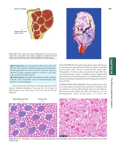
847


           Figure 28.17  Giant cell tumour (osteoclastoma). The end of the
           long bone is expanded in the region of epiphysis. Sectioned surface
           shows circumscribed, dark tan, haemorrhagic and necrotic tumour.


               Stromal cells are mononuclear cells and are the real  CELL OF ORIGIN. Though designated as giant cell tumour  CHAPTER 28
            tumour cells and their histologic appearance determines  or osteoclastoma, the true tumour cells are round to spindled
            the biologic behaviour of the tumour. Typically, they are  mononuclear cells and not osteoclast-like giant cells.
            uniform, plump, spindle-shaped or round to oval cells  Histogenesis of tumour cells is uncertain but possibly they
            with numerous mitotic figures.                     are of mesenchymal origin. Available evidence suggests that
               Other features of the stroma include its scanty collagen  osteoclasts are derived from fusion of circulating monocytes,
            content, rich vascularity, areas of haemorrhages and  the process being facilitated by transforming growth factor-
            presence of macrophages.                           beta (TGF-β).
                                                               OTHER GIANT CELL LESIONS. This peculiar tumour with
              Giant cell tumour of bone has certain peculiarities which  above description is named ‘giant cell tumour’ but giant cells
           deserve further elaboration. These are: its cell of origin, its  are present in several other benign tumours and tumour-
           differentiation from other giant cell lesions and its  biologic  like lesions from which the giant cell tumour is to be
           behaviour.                                                                                                 The Musculoskeletal System
                                                               distinguished. These  benign giant cell lesions are:


           Figure 28.18  Osteoclastoma. Microscopy reveals osteoclast-like multinucleate giant cells which are regularly distributed among the mononuclear
           stromal cells.