
Page 866 - Textbook of Pathology, 6th Edition
P. 866
850 composed of inner layer of 1-4 cell thick synoviocytes and
outer layer of loose vascular connective tissue. On electron
microscopy, two types of synoviocytes are distinguished:
type A and type B. Type A synoviocytes are more numerous
and are related to macrophages and produce degradative
enzymes, while type B synthesise hyaluronic acid.
Diseases of joints are numerous and joints are also invol-
ved in several systemic disorders. In the following discussion,
only those joint diseases which are morphologically
significant are described. Synovial tumours are discussed in
the next chapter together with other soft tissue tumours.
OSTEOARTHRITIS
Osteoarthritis (OA), also called osteoarthrosis or degenerative
joint disease (DJD), is the most common form of chronic
disorder of synovial joints. It is characterised by progressive
degenerative changes in the articular cartilages over the
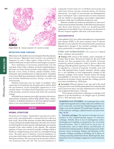
Figure 28.22 Osseous deposits from carcinoma breast. years, particularly in weight-bearing joints.
METASTATIC BONE TUMOURS TYPES AND PATHOGENESIS. OA occurs in 2 clinical
forms—primary and secondary.
Metastases to the skeleton are more frequent than the primary Primary OA occurs in the elderly, more commonly in
bone tumours. Metastatic bone tumours are exceeded in women than in men. The process begins by the end of 4th
frequency by only 2 other organs—lungs and liver. Most decade and then progressively and steadily increases
skeletal metastases are derived from haematogenous spread. producing clinical symptoms. Little is known about the
Bony metastases of carcinomas predominate over the etiology and pathogenesis of primary OA. The condition may
sarcomas. Some of the common carcinomas metastasising to be regarded as a reward of longevity. Probably, wear and
the bones are from: breast, prostate, lung, kidney, stomach, tear with repeated minor trauma, heredity, obesity, aging
thyroid, cervix, body of uterus, urinary bladder, testis, per se, all contribute to focal degenerative changes in the
SECTION III
melanoma and neuroblastoma of adrenal gland. Examples articular cartilage of the joints. Genetic factors favouring
of sarcomas which may metastasise to the bone are: embryonal susceptibility to develop OA have been observed; genetic
and alveolar rhabdomyosarcoma, Ewing’s sarcoma and mutations in proteins which regulate the cartilage growth
osteosarcoma. have been identified e.g. FRZB gene.
Skeletal metastases may be single or multiple. Most
Secondary OA may appear at any age and is the result
commonly involved bones are: the spine, pelvis, femur, skull, of any previous wear and tear phenomena involving the joint
ribs and humerus. Usual radiographic appearance is of an
osteolytic lesion. Osteoblastic bone metastases occur in cancer such as previous injury, fracture, inflammation, loose bodies
of the prostate, carcinoid tumour and small cell carcinoma and congenital dislocation of the hip.
of lung. The molecular mechanism of damage to cartilage in OA
Metastatic bone tumours generally reproduce the micros- appears to be the breakdown of collagen type II, probably
Systemic Pathology
copic picture of primary tumour (Fig. 28.22). Many a times, by IL-1, TNF and nitric oxide.
evidence of skeletal metastases is the first clinical manifes- MORPHOLOGIC FEATURES. As mentioned above, the
tation of an occult primary cancer in the body. weight-bearing joints such as hips, knee and vertebrae are
most commonly involved but interphalangeal joints of
JOINTS fingers may also be affected. The pathologic changes occur
in the articular cartilages, adjacent bones and synovium
NORMAL STRUCTURE (Fig. 28.23):
The joints are of 2 types—diarthrodial or synovial joints with a 1. Articular cartilages. The regressive changes are most
joint cavity, and synarthrodial or nonsynovial joints without a marked in the weight-bearing regions of articular
joint cavity. Most of the diseases of joints affect diarthrodial cartilages. Initially, there is loss of cartilaginous matrix
or synovial joints. In diarthrodial joints, the ends of two bones (proteoglycans) resulting in progressive loss of normal
are held together by joint capsule with ligaments and tendons metachromasia. This is followed by focal loss of
inserted at the outer surface of the capsule. The articular chondrocytes, and at other places, proliferation of
surfaces of bones are covered by hyaline cartilage which is chondrocytes forming clusters. Further progression of the
thicker in weight-bearing areas than in nonweight-bearing process causes loosening, flaking and fissuring of the
areas. The joint space is lined by synovial membrane or articular cartilage resulting in breaking off of pieces of
synovium which forms synovial fluid that lubricates the joint cartilage exposing subchondral bone. Radiologically, this
during movements. The synovium may be smooth or thrown progressive loss of cartilage is apparent as narrowed joint
into numerous folds and villi. The synovial membrane is space.