
Page 577 - First Aid for the USMLE Step 1 2020, Thirtieth edition [MedicalBooksVN.com]_Neat
P. 577
Neurology aNd Special SeNSeS ` neurology—PAthology Neurology aNd Special SeNSeS ` neurology—otology SecTioN iii 533
` neurology—otology
Auditory physiology
Outer ear Visible portion of ear (pinna), includes auditory canal and tympanic membrane. Transfers sound
waves via vibration of tympanic membrane.
Middle ear Air-filled space with three bones called the ossicles (malleus, incus, stapes). Ossicles conduct and
amplify sound from tympanic membrane to inner ear.
Inner ear Snail-shaped, fluid-filled cochlea. Contains basilar membrane that vibrates 2° to sound waves.
Vibration transduced via specialized hair cells auditory nerve signaling brain stem.
Each frequency leads to vibration at specific location on basilar membrane (tonotopy):
Low frequency heard at apex near helicotrema (wide and flexible).
High frequency heard best at base of cochlea (thin and rigid).
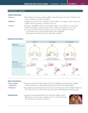
Diagnosing hearing loss
Normal Conductive Sensorineural
Weber test
Tuning fork on vertex of skull
Localizes to a ected ear Localizes to una ected ear
No localization
↓ transmission of background noise ↓ transmission of all sound
Rinne test
Tuning fork in front of ear
(air conduction, AC),
Tuning fork on mastoid
process (bone conduction, BC)
AC > BC BC > AC AC > BC
Types of hearing loss
Noise-induced Damage to stereociliated cells in organ of Corti. Loss of high-frequency hearing first. Sudden
hearing loss extremely loud noises can produce hearing loss due to tympanic membrane rupture.
Presbycusis Aging-related progressive bilateral/symmetric sensorineural hearing loss (often of higher frequencies)
due to destruction of hair cells at the cochlear base (preserved low-frequency hearing at apex).
Cholesteatoma Overgrowth of desquamated keratin debris within the middle ear space A
( A , arrows); may erode ossicles, mastoid air cells conductive hearing
loss. Often presents with painless otorrhea.
FAS1_2019_12-Neurol.indd 533 11/8/19 7:39 AM