
Page 741 - First Aid for the USMLE Step 1 2020, Thirtieth edition [MedicalBooksVN.com]_Neat
P. 741
Rapid Review ` ClassiC labs/FinDinGs Rapid Review ` ClassiC labs/FinDinGs SeCTiON iii 697
lab/DiaGnostiC FinDinG DiaGnosis/Disease PaGe
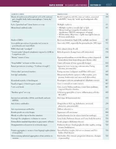
Sheets of medium-sized lymphoid cells with scattered Burkitt lymphoma (t[8:14] c-myc activation, associated 430
pale, tingible body–laden macrophages (“starry sky” with EBV; “starry sky” made up of malignant cells)
histology)
Lytic (“punched-out”) bone lesions on x-ray Multiple myeloma 431
Monoclonal antibody spike Multiple myeloma (usually IgG or IgA) 431
Monoclonal gammopathy of undetermined
significance (MGUS consequence of aging)
Waldenström (M protein = IgM) macroglobulinemia
Primary amyloidosis
Stacks of RBCs Rouleaux formation (high ESR, multiple myeloma) 423
Azurophilic peroxidase ⊕ granular inclusions in Auer rods (AML, especially the promyelocytic [M3] type) 432
granulocytes and myeloblasts
WBCs that look “smudged” CLL (almost always B cell) 432
“Tennis racket”-shaped cytoplasmic organelles (EM) in Birbeck granules (Langerhans cell histiocytosis) 434
Langerhans cells
“Brown” tumor of bone Hyperparathyroidism or osteitis fibrosa cystica (deposited 464
hemosiderin from hemorrhage gives brown color)
“Soap bubble” in femur or tibia on x-ray Giant cell tumor of bone (generally benign) 464
Raised periosteum (creating a “Codman triangle”) Aggressive bone lesion (eg, osteosarcoma, Ewing 465
sarcoma, osteomyelitis)
“Onion skin” periosteal reaction Ewing sarcoma (malignant small blue cell tumor) 465
Anti-IgG antibodies Rheumatoid arthritis (systemic inflammation, joint 466
pannus, boutonniere and swan neck deformities)
Rhomboid crystals, ⊕ birefringent Pseudogout (calcium pyrophosphate dihydrate crystals) 467
Needle-shaped, ⊝ birefringent crystals Gout (monosodium urate crystals) 467
uric acid levels Gout, Lesch-Nyhan syndrome, tumor lysis syndrome, 467
loop and thiazide diuretics
“Bamboo spine” on x-ray Ankylosing spondylitis (chronic inflammatory arthritis: 469
HLA-B27)
Antinuclear antibodies (ANAs: anti-Smith and anti- SLE (type III hypersensitivity) 470
dsDNA)
Anti-histone antibodies Drug-induced SLE (eg, hydralazine, isoniazid, 250
phenytoin, procainamide)
Anti-topoisomerase antibodies Diffuse scleroderma 473
Keratin pearls on a skin biopsy Squamous cell carcinoma 484
Bloody or yellow tap on lumbar puncture Xanthochromia (due to subarachnoid hemorrhage) 513
Eosinophilic cytoplasmic inclusion in neuron Lewy body (Parkinson disease and Lewy body dementia) 520
Extracellular amyloid deposition in gray matter of brain Senile plaques (Alzheimer disease) 520
Depigmentation of neurons in substantia nigra Parkinson disease (basal ganglia disorder: rigidity, resting 520
tremor, bradykinesia)
Protein aggregates in neurons from hyperphosphorylation Neurofibrillary tangles (Alzheimer disease) and Pick 520
of tau protein bodies (Pick disease)
Silver-staining spherical aggregation of tau proteins in Pick bodies (Pick disease: progressive dementia, changes 520
neurons in personality)
FAS1_2019_17_Rapid Rev.indd 697 11/7/19 6:09 PM