
Page 100 - The Netter Collection of Medical Illustrations - Integumentary System_ Volume 4 ( PDFDrive )
P. 100
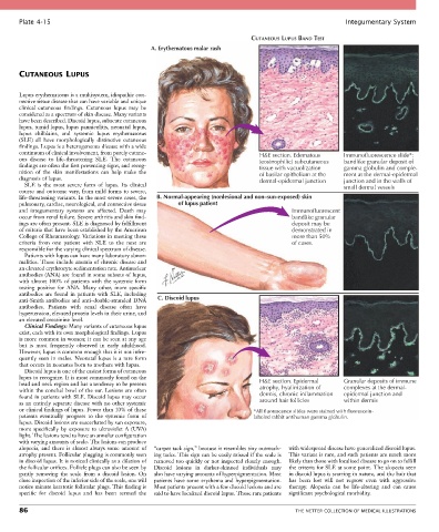
Plate 4-15                                                                                            Integumentary System

                                                                               CUTANEOUS LUPUS BAND TEST
                                                A. Erythematous malar rash


       CUTANEOUS LUPUS


       Lupus erythematosus is a multisystem, idiopathic con-
       nective tissue disease that can have variable and unique
       clinical  cutaneous  findings.  Cutaneous  lupus  may  be
       considered as a spectrum of skin disease. Many variants
       have been described. Discoid lupus, subacute cutaneous
       lupus, tumid lupus, lupus panniculitis, neonatal lupus,
       lupus  chilblains,  and  systemic  lupus  erythematosus
       (SLE)  all  have  morphologically  distinctive  cutaneous
       findings. Lupus is a heterogeneous disease with a wide
       continuum of clinical involvement, from purely cutane-                     H&E section. Edematous    Immunofluorescence slide*:
       ous  disease  to  life-threatening  SLE.  The  cutaneous                   (eosinophilic) subcutaneous  bandlike granular deposit of
       findings are often the first presenting signs, and recog-                  tissue with vacuolization  gamma globulin and comple-
       nition  of  the  skin  manifestations  can  help  make  the                of basilar epithelium at the  ment at the dermal-epidermal
       diagnosis of lupus.                                                        dermal-epidermal junction  junction and in the walls of
         SLE  is  the  most  severe  form  of  lupus.  Its  clinical                                        small dermal vessels
       course and outcome vary, from mild forms to severe,
       life-threatening variants. In the most severe cases, the   B. Normal-appearing (nonlesional and non–sun-exposed) skin
       pulmonary, cardiac, neurological, and connective tissue   of lupus patient
       and  integumentary  systems  are  affected.  Death  may                              Immunofluorescent
       occur from renal failure. Severe arthritis and skin find-                            bandlike granular
       ings are often present. SLE is diagnosed by fulfillment                              deposit may be
       of criteria that have been established by the American                               demonstrated in
       College of Rheumatology. Variations in meeting these                                 more than 50%
       criteria  from  one  patient  with  SLE  to  the  next  are                          of cases.
       responsible for the varying clinical spectrum of disease.
         Patients with lupus can have many laboratory abnor-
       malities. These include anemia of chronic disease and
       an elevated erythrocyte sedimentation rate. Antinuclear
       antibodies (ANA) are found in some subsets of lupus,
       with almost 100% of patients with the systemic form
       testing  positive  for  ANA.  Many  other,  more  specific
       antibodies  are  found  in  patients  with  SLE,  including
       anti-Smith antibodies and anti–double-stranded DNA   C. Discoid lupus
       antibodies.  Patients  with  renal  disease  often  have
       hypertension, elevated protein levels in their urine, and
       an elevated creatinine level.
         Clinical Findings: Many variants of cutaneous lupus
       exist, each with its own morphological findings. Lupus
       is more common in women; it can be seen at any age
       but  is  most  frequently  observed  in  early  adulthood.
       However, lupus is common enough that it is not infre-
       quently  seen  in  males.  Neonatal  lupus  is  a  rare  form
       that occurs in neonates born to mothers with lupus.
         Discoid lupus is one of the easiest forms of cutaneous
       lupus to recognize. It is most commonly found on the
       head and neck region and has a tendency to be present                      H&E section. Epidermal    Granular deposits of immune
       within the conchal bowl of the ear. Lesions are often                      atrophy, hyalinization of  complexes at the dermal-
       found in patients with SLE. Discoid lupus may occur                        dermis, chronic inflammation   epidermal junction and
       as an entirely separate disease with no other systemic                     around hair follicles     within dermis
       or clinical findings of lupus. Fewer than 10% of these                   *All fluorescence slides were stained with fluorescein-
       patients  eventually  progress  to  the  systemic  form  of              labeled rabbit antihuman gamma globulin.
       lupus. Discoid lesions are exacerbated by sun exposure,
       more  specifically  by  exposure  to  ultraviolet  A  (UVA)
       light. The lesions tend to have an annular configuration
       with varying amounts of scale. The lesions can produce
       alopecia,  and  there  is  almost  always  some  amount  of   “carpet tack sign,” because it resembles tiny outreach-  with widespread disease have generalized discoid lupus.
       atrophy present. Follicular plugging is commonly seen   ing tacks. This sign can be easily missed if the scale is   This variant is rare, and such patients are much more
       in discoid lupus. It is noticed clinically as a dilation of   removed too quickly or not inspected closely enough.   likely than those with localized disease to go on to fulfill
       the follicular orifices. Follicle plugs can also be seen by   Discoid  lesions  in  darker-skinned  individuals  may     the criteria for SLE at some point. The alopecia seen
       gently  removing  the  scale  from  a  discoid  lesion.  On   also have varying amounts of hyperpigmentation. Most   in discoid lupus is scarring in nature, and the hair that
       close inspection of the inferior side of the scale, one will   patients have some erythema and hyperpigmentation.   has  been  lost  will  not  regrow  even  with  aggressive
       notice minute keratotic follicular plugs. This finding is   Most patients present with a few discoid lesions and are   therapy.  Alopecia  can  be  life-altering  and  can  cause
       specific  for  discoid  lupus  and  has  been  termed  the   said to have localized discoid lupus. Those rare patients   significant psychological morbidity.

       86                                                                                    THE NETTER COLLECTION OF MEDICAL ILLUSTRATIONS