
Page 104 - The Netter Collection of Medical Illustrations - Integumentary System_ Volume 4 ( PDFDrive )
P. 104
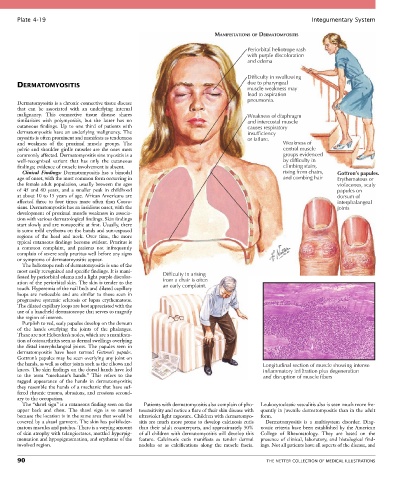
Plate 4-19                                                                                            Integumentary System

                                                                           MANIFESTATIONS OF DERMATOMYOSITIS

                                                                                      Periorbital heliotrope rash
                                                                                      with purple discoloration
                                                                                      and edema


                                                                                      Difficulty in swallowing
       DERMATOMYOSITIS                                                                due to pharyngeal
                                                                                      muscle weakness may
                                                                                      lead to aspiration
                                                                                      pneumonia.
       Dermatomyositis is a chronic connective tissue disease
       that  can  be  associated  with  an  underlying  internal
       malignancy.  This  connective  tissue  disease  shares                         Weakness of diaphragm
       similarities  with  polymyositis,  but  the  latter  has  no                   and intercostal muscle
       cutaneous  findings.  Up  to  one  third  of  patients  with                   causes respiratory
       dermatomyositis have an underlying malignancy. The                             insufficiency
       myositis is often prominent and manifests as tenderness                        or failure.
       and  weakness  of  the  proximal  muscle  groups.  The                                      Weakness of
       pelvic and shoulder girdle muscles are the ones most                                        central muscle
       commonly affected. Dermatomyositis sine myositis is a                                       groups evidenced
       well-recognized  variant  that  has  only  the  cutaneous                                   by difficulty in
       findings; evidence of muscle involvement is absent.                                         climbing stairs,
         Clinical  Findings:  Dermatomyositis  has  a  bimodal                                     rising from chairs,  Gottron’s papules.
       age of onset, with the most common form occurring in                                        and combing hair  Erythematous or
       the female adult population, usually between the ages                                                         violaceous, scaly
       of  45  and  60  years,  and  a  smaller  peak  in  childhood                                                 papules on
       at about 10 to 15 years of age. African Americans are                                                         dorsum of
       affected  three  to  four  times  more  often  than  Cauca-                                                   interphalangeal
       sians. Dermatomyositis has an insidious onset, with the                                                       joints
       development of proximal muscle weakness in associa-
       tion with various dermatological findings. Skin findings
       start slowly and are nonspecific at first. Usually, there
       is some mild erythema on the hands and sun-exposed
       regions  of  the  head  and  neck.  Over  time,  the  more
       typical cutaneous findings become evident. Pruritus is
       a  common  complaint,  and  patients  not  infrequently
       complain of severe scalp pruritus well before any signs
       or symptoms of dermatomyositis appear.
         The heliotrope rash of dermatomyositis is one of the
       most easily recognized and specific findings. It is mani-  Difficulty in arising
       fested by periorbital edema and a light purple discolor-  from a chair is often
       ation of the periorbital skin. The skin is tender to the   an early complaint.
       touch. Hyperemia of the nail beds and dilated capillary
       loops  are  noticeable  and  are  similar  to  those  seen  in
       progressive  systemic  sclerosis  or  lupus  erythematous.
       The dilated capillary loops are best appreciated with the
       use of a handheld dermatoscope that serves to magnify
       the region of interest.
         Purplish to red, scaly papules develop on the dorsum
       of  the  hands  overlying  the  joints  of  the  phalanges.
       These are not Heberden’s nodes, which are a manifesta-
       tion of osteoarthritis seen as dermal swellings overlying
       the  distal  interphalangeal  joints.  The  papules  seen  in
       dermatomyositis  have  been  termed  Gottron’s  papules.
       Gottron’s papules may be seen overlying any joint on
       the hands, as well as other joints such as the elbows and                            Longitudinal section of muscle showing intense
       knees. The skin findings on the dorsal hands have led                                inflammatory infiltration plus degeneration
       to  the  term  “mechanic’s  hands.”  This  refers  to  the                           and disruption of muscle fibers
       ragged  appearance  of  the  hands  in  dermatomyositis;
       they resemble the hands of a mechanic that have suf-
       fered chronic trauma, abrasions, and erosions second-
       ary to the occupation.
         The “shawl sign” is a cutaneous finding seen on the   Patients with dermatomyositis also complain of pho-  Leukocytoclastic vasculitis also is seen much more fre-
       upper  back  and  chest.  The  shawl  sign  is  so  named   tosensitivity and notice a flare of their skin disease with   quently in juvenile dermatomyositis than in the adult
       because the location is in the same area that would be   ultraviolet light exposure. Children with dermatomyo-  form.
       covered by a shawl garment. The skin has poikiloder-  sitis are much more prone to develop calcinosis cutis   Dermatomyositis  is  a  multisystem  disorder.  Diag-
       matous macules and patches. There is a varying amount   than their adult counterparts, and approximately 50%   nostic criteria have been established by the American
       of skin atrophy with telangiectases, mottled hyperpig-  of all children with dermatomyositis will develop this   College  of  Rheumatology.  They  are  based  on  the
       mentation and hypopigmentation, and erythema of the   feature.  Calcinosis  cutis  manifests  as  tender  dermal   presence  of  clinical,  laboratory,  and  histological  find-
       involved region.                          nodules  or  as  calcifications  along  the  muscle  fascia.   ings. Not all patients have all aspects of the disease, and

       90                                                                                    THE NETTER COLLECTION OF MEDICAL ILLUSTRATIONS