
Page 105 - The Netter Collection of Medical Illustrations - Integumentary System_ Volume 4 ( PDFDrive )
P. 105
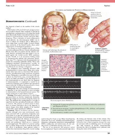
Plate 4-20                                                                                                           Rashes

                                                                   CUTANEOUS AND LABORATORY FINDINGS IN DERMATOMYOSITIS
                                                                                                             Head in flexed
                                                                                                             position due to
                                                                                                             proximal muscle
                                                                                                             weakness

        DERMATOMYOSITIS (Continued)


        the  diagnosis  is  based  on  the  number  of  the  criteria
        fulfilled.
          Inflammation of the proximal muscle groups has been
        well described. Patients often complain of difficulty in
        standing from a sitting position or in raising their hands
        above their heads. Patients have elevated serum concen-
        trations of creatinine kinase, aldolase, and lactate dehy-
        drogenase.  This  is  indicative  of  muscle  inflammation
        and  breakdown.  An  electromyogram  (EMG)  can  be
        used  to  evaluate  the  weakness  and  to  differentiate  a
        nerve  origin  from  a  muscle  origin.  A  muscle  biopsy,
        most  commonly  of  the  deltoid  muscle,  shows  active                    Difficulty in
        inflammation on histological examination.                                   swallowing due
          This disease can rarely manifest with severe, diffuse                     to esophageal                 Gottron’s papules.
        interstitial pulmonary fibrosis. Patients with pulmonary   Edema and heliotrope discoloration  weakness   Erythematous, nodular
        fibrosis most often test positive for the anti-Jo1 anti-  of eyelids; erythematous rash                   eruption on fingers
        body. Anti-Jo1 antibodies have been found to be tar-
        geted  against  the  histidyl–transfer  RNA  synthetase
        protein.  Overall,  it  is  an  uncommon  finding  except
        in  dermatomyositis  patients  with  pulmonary  disease.
        More than 75% of patients with dermatomyositis test   Atrophy                                                    Immuno-
        positive for antinuclear antibodies (ANA). Those with   of muscle                                                globulin
        malignancy-associated  dermatomyositis  typically  do   fibers and                                               deposition
        not develop pulmonary fibrosis, and those with pulmo-  lymphocyte                                                in blood
        nary fibrosis do not develop a malignancy.  infiltration                                                         vessel of
          The  malignancy  most  commonly  associated  with   (muscle                                                    muscle
        dermatomyositis is ovarian cancer. Many other malig-  biopsy)                                                    (immuno-
        nancies  have  been  seen  in  association  with  dermato-                                                       fluorescence)
        myositis, including breast, lung, lymphoma, and gastric
        cancers. Malignancy is seen before the onset of the rash
        in about one third of the cases, concurrently with the
        rash in one third, and within 2 years after diagnosis of
        the dermatomyositis in one third. After the diagnosis
        of  dermatomyositis,  it  is  imperative  to  search  for  an
        underlying malignancy and to perform age-appropriate   Normal
        cancer screening. Childhood dermatomyositis is rarely
        associated with an underlying malignancy.
          Pathogenesis: The exact etiology of dermatomyositis
        is unknown. It has been theorized to occur secondary
        to abnormalities in the humoral immune system. The
        precise mechanism is under intense research.  Myopathy
          Histology: Histological examination of a skin biopsy
        specimen  shows  an  interface  lymphocytic  dermatitis.
        Hydropic change is seen scattered along the basilar cell
        layer. The epidermis has varying degrees of atrophy. A   Electromyogram shows fibrillations
        superficial and deep periadnexal lymphocytic infiltrate
        is  common.  The  presence  of  dermal  mucin  in  abun-  1. Nonspecific hypergammaglobulinemia; low incidence of antinuclear antibodies
        dance  is  another  histological  clue  to  the  diagnosis.  A        and rheumatoid factor
        muscle  biopsy  often  shows  atrophy  of  the  involved   Laboratory
        muscle with a dense lymphocytic infiltrate.  findings  2. Elevated serum enzymes. creatine phosphokinase (CPK), aldolase, and aspartate
          Treatment:  There  is  no  known  cure  for  dermato-      amine transferase (AST, SGOT)
        myositis,  although  some  cases  spontaneously  remit.   3. Elevated urinary creatine and myoglobulin levels
        Cases associated with an underlying malignancy have
        been shown to go into full remission with cure of the
        underlying cancer. Relapse of dermatomyositis in these
        patients  should  prompt  the  clinician  to  search  for  a   agent to keep the disease at bay. Many steroid-sparing   the  itching  and  decrease  some  of  the  redness.  The
        recurrence  of  their  malignancy.  Initial  treatment  is   agents have been used, including hydroxychloroquine,   treatment  of  juvenile  dermatomyositis  is  similar.  It  is
        usually  with  prednisone,  which  acts  as  a  nonspecific   quinacrine, cyclosporine, intravenous immunoglobulin   believed to have a better prognosis, because few cases
        immunosuppressant. The addition of a steroid-sparing   (IVIG), azathioprine, and methotrexate, all with vari-  are associated with an underlying cancer. It is thought
        agent is almost always needed to avoid the long-term   able success. Combination therapy is the norm.  that  early  treatment  of  juvenile  dermatomyositis
        side  effects  of  prednisone.  Some  patients  require  a   The  use  of  sun  protection  and  sunscreen  cannot     decreases the risk of developing severe calcinosis cutis
        smaller dose of prednisone along with a steroid-sparing   be overemphasized. Topical corticosteroids help relieve   during the course of the disease.


        THE NETTER COLLECTION OF MEDICAL ILLUSTRATIONS                                                                           91