
Page 101 - The Netter Collection of Medical Illustrations - Integumentary System_ Volume 4 ( PDFDrive )
P. 101
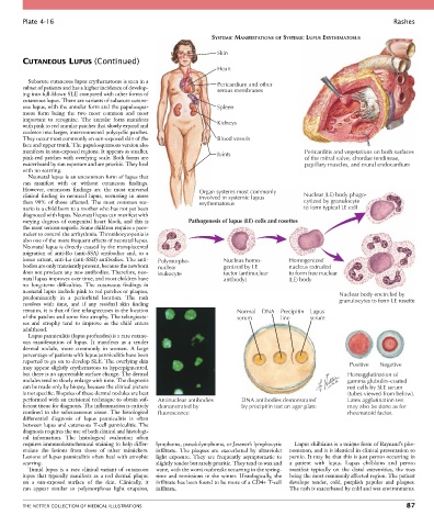
Plate 4-16                                                                                                           Rashes

                                                                    SYSTEMIC MANIFESTATIONS OF SYSTEMIC LUPUS ERYTHEMATOSUS

                                                                     Skin
        CUTANEOUS LUPUS (Continued)
                                                                     Heart
          Subacute cutaneous lupus erythematosus is seen in a
        subset of patients and has a higher incidence of develop-    Pericardium and other
                                                                     serous membranes
        ing into full-blown SLE compared with other forms of
        cutaneous lupus. There are variants of subacute cutane-
        ous lupus, with the annular form and the papulosqua-         Spleen
        mous  form  being  the  two  most  common  and  most
        important  to  recognize.  The  annular  form  manifests
        with pink to red annular patches that slowly expand and      Kidneys
        coalesce into larger, interconnected polycyclic patches.
        They occur most commonly on sun-exposed skin of the          Blood vessels
        face and upper trunk. The papulosquamous version also
        manifests in sun-exposed regions. It appears as smaller,     Joints                      Pericarditis and vegetations on both surfaces
        pink-red patches with overlying scale. Both forms are                                    of the mitral valve, chordae tendineae,
        exacerbated by sun exposure and are pruritic. They heal                                  papillary muscles, and mural endocardium
        with no scarring.
          Neonatal lupus is an uncommon form of lupus that
        can  manifest  with  or  without  cutaneous  findings.
        However,  cutaneous  findings  are  the  most  universal   Organ systems most commonly
        clinical  finding  in  neonatal  lupus,  occurring  in  more   involved in systemic lupus  Nuclear (LE) body phago-
        than  90%  of  those  affected.  The  most  common  sce-  erythematosus                  cytized by granulocyte
        nario is a child born to a mother who has not yet been                                   to form typical LE cell
        diagnosed with lupus. Neonatal lupus can manifest with
        varying degrees of congenital heart block, and this is   Pathogenesis of lupus (LE) cells and rosettes
        the most serious sequela. Some children require a pace-
        maker to control the arrhythmia. Thrombocytopenia is
        also one of the more frequent effects of neonatal lupus.
        Neonatal lupus is directly caused by the transplacental
        migration  of  anti-Ro  (anti-SSA)  antibodies  and,  to  a
        lesser extent, anti-La (anti-SSB) antibodies. The anti-  Polymorpho-  Nucleus homo-  Homogenized
        bodies are only transiently present, because the newborn   nuclear  genized by LE   nucleus extruded
        does not produce any new antibodies. Therefore, neo-  leukocyte  factor (antinuclear  to form free nuclear
        natal lupus improves over time, and most children have         antibody)            (LE) body
        no  long-term  difficulties.  The  cutaneous  findings  in
        neonatal lupus include pink to red patches or plaques,                                              Nuclear body encircled by
        predominantly  in  a  periorbital  location.  The  rash                                             granulocytes to form LE rosette
        resolves  with  time,  and  if  any  residual  skin  finding
        remains, it is that of fine telangiectases in the location          Normal  DNA  Precipitin  Lupus
        of the patches and some fine atrophy. The telangiecta-              serum        line      serum
        ses  and  atrophy  tend  to  improve  as  the  child  enters
        adulthood.
          Lupus panniculitis (lupus profundus) is a rare cutane-
        ous  manifestation  of  lupus.  It  manifests  as  a  tender
        dermal  nodule,  more  commonly  in  women.  A  large
        percentage of patients with lupus panniculitis have been
        reported to go on to develop SLE. The overlying skin                                                   Positive  Negative
        may appear slightly erythematous to hyperpigmented,
        but there is no appreciable surface change. The dermal                                                 Hemagglutination of
        nodules tend to slowly enlarge with time. The diagnosis                                                gamma globulin–coated
        can be made only by biopsy, because the clinical picture                                               red cells by SLE serum
        is not specific. Biopsies of these dermal nodules are best                                             (tubes viewed from below).
        performed with an excisional technique to obtain suf-  Antinuclear antibodies  DNA antibodies demonstrated   Latex agglutination test
        ficient tissue for diagnosis. The inflammation is entirely   demonstrated by  by precipitin test on agar plate  may also be done as for
        confined  to  the  subcutaneous  tissue.  The  histological   fluorescence                             rheumatoid factor.
        differential  diagnosis  of  lupus  panniculitis  is  often
        between lupus and cutaneous T-cell panniculitis. The
        diagnosis requires the use of both clinical and histologi-
        cal  information.  The  histological  evaluation  often
        requires immunohistochemical staining to help differ-  lymphoma, pseudolymphoma, or Jessner’s lymphocytic   Lupus chilblains is a unique form of Raynaud’s phe-
        entiate  the  lesions  from  those  of  other  mimickers.   infiltrate.  The  plaques  are  exacerbated  by  ultraviolet   nomenon, and it is identical in clinical presentation to
        Lesions of lupus panniculitis often heal with atrophic   light  exposure.  They  are  frequently  asymptomatic  to   pernio. It may be that this is just pernio occurring in
        scarring.                                 slightly tender but rarely pruritic. They tend to wax and   a  patient  with  lupus.  Lupus  chilblains  and  pernio
          Tumid  lupus  is  a  rare  clinical  variant  of  cutaneous   wane, with the worst outbreaks occurring in the spring-  manifest  typically  on  the  distal  extremities,  the  toes
        lupus  that  typically  manifests  as  a  red  dermal  plaque     time and remissions in the winter. Histologically, the   being the most commonly affected region. The patient
        on  a  sun-exposed  surface  of  the  skin.  Clinically,  it     infiltrate has been found to be more of a CD4+ T-cell   develops  tender,  cold,  purplish  papules  and  plaques.
        can  appear  similar  to  polymorphous  light  eruption,   infiltrate.              The rash is exacerbated by cold and wet environments.


        THE NETTER COLLECTION OF MEDICAL ILLUSTRATIONS                                                                           87