
Page 99 - The Netter Collection of Medical Illustrations - Integumentary System_ Volume 4 ( PDFDrive )
P. 99
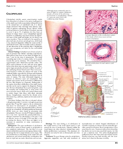
Plate 4-14                                                                                                           Rashes

                                                     Although most commonly seen in
                                                     areas of high fat content (abdomen,
        CALCIPHYLAXIS                                breast), all areas of the skin may
                                                     be involved in calciphylaxis. Almost
                                                     all cases are associated with
        Calciphylaxis  (calcific  uremic  arteriolopathy)  results   underlying renal disease.
        from deposition of calcium in the tunica media portion
        of the small vessel walls in association with proliferation
        of  the  intimal  layer  of  endothelial  cells.  It  is  almost
        always  associated  with  end-stage  renal  disease,  espe-
        cially  in  patients  undergoing  chronic  dialysis  (either
        peritoneal dialysis or hemodialysis). It has been reported
        to  occur  in  up  to  5%  of  patients  who  have  been  on
        dialysis for longer than 1 year. Calciphylaxis typically
        manifests as nonhealing skin ulcers located in adipose-                                 Calcium deposits in the conduction system
        rich areas of the trunk and thighs, but the lesions can                                 of the heart, which may cause serious
        occur anywhere. They are believed to be caused by an                                    or fatal arrhythmias
        abnormal ratio of calcium and phosphorus, which leads
        to the abnormal deposition within the tunica media of
        small blood vessels. This eventually results in thrombo-
        sis and ulceration of the overlying skin. Calciphylaxis
        has  a  poor  prognosis,  and  there  are  few  well-studied
        therapies.
          Clinical Findings: Calciphylaxis is almost exclusively
        seen in patients with chronic end-stage renal disease.
        Most patients have been on one form of dialysis for at
        least  1  year  by  the  time  of  presentation.  The  initial
        presenting sign is that of a tender, dusky red to purple
        macule that quickly ulcerates. The ulcerations have a
        ragged border and a thick black necrotic eschar. The                                    Medial calcification of small arteries and
        ulcers  tend  to  increase  in  size,  and  new  areas  appear                          arterioles
        before older ulcers have any opportunity to heal. Ulcer-
        ations begin proximally and tend to follow the path of                          Avascular
        the underlying affected blood vessel. Their most prom-                            zone
        inent  location  is  within  the  adipose-rich  areas  of  the
        trunk and thighs, especially the abdomen and mammary                                                    Endothelium
        regions. Patients often report that ulcerations form in                                                 Desmosome
        areas  of  trauma.  The  main  differential  diagnosis  is                                              Basement membrane
        between  an  infectious  cause  and  calciphylaxis.  Skin                  Vascular                    Smooth muscle    Intima
        biopsies and cultures can be performed to differentiate                     zone                       cell (atherophil)
        the two. Skin biopsies are diagnostic. Radiographs of                                                  Fibroblast  Lamina
        the region often show calcification of the small vessels,                                              Collagen   propria
                                                                                                               Matrix
        and this can be used to support the diagnosis. Patients                                                      Internal elastic
        who develop calciphylaxis have a poor prognosis, with                                                        membrane
        the  mortality  rate  reaching  80%  in  some  series.  For                                                  Muscle and  Media
                                                                                                                     elastic tissue
        some unknown reason, those with truncal disease tend                                                         Reticular fibers
        to  survive  longer  than  those  with  distal  extremity                                                    External elastic
                                                                                                                     membrane
        disease.  Complications  caused  by  the  chronic  severe
        ulcerations (e.g., infection, sepsis) are the main cause of                                                    Adventitia
        mortality.
          Laboratory findings often show an elevated calcium
        × phosphorus product. A calcium × phosphorus product                                                           Vasa vasorum
                         2
                      2
        greater than 70 mg /dL  appears to be an independent
        risk  factor  for  development  of  calciphylaxis.  Other
        risk factors are obesity, hyperparathyroidism, diabetes,
        and the use of warfarin. Elevated parathyroid hormone
        (PTH)  levels  are  often  found  in  association  with
        calciphylaxis. The exact role that PTH plays is unknown,                                                      Sympathetic nerve
        but  it  has  been  reported  that  parathyroidectomy,  a                                                     (vasomotor)
        standard treatment for calciphylaxis in the past, is not                        Wall of an artery: cutaway view
        an  effective  means  of  therapy.  PTH  may  play  a  role
        in  starting  the  disease,  but  it  does  not  appear  to  be
        necessary  to  exacerbate  or  cause  continuation  of
        calciphylaxis.                              Histology:  The  main  finding  is  of  calcification  of   superinfections  are  critical.  Surgical  debridement  of
          Pathogenesis: The exact mechanism of calcification   the  medial  section  of  the  small  blood  vessels  in  and   wounds is necessary to remove necrotic tissue that pro-
        of the tunica media of blood vessels in calciphylaxis is   around  the  area  of  involvement.  Thrombi  within  the   vides a portal for infection. Renal transplantation offers
        not  completely  understood.  The  fact  that  it  is  seen   vessel  lumen  are  often  observed.  Intimal  layer  endo-  some hope for cure. Treatment with sodium thiosulfate
        almost exclusively in patients undergoing chronic dialy-  thelial  proliferation  is  prominent.  The  abnormal  cal-  has shown success in some anecdotal reports, but this
        sis therapy has led to many theories on its origin. The   cification can easily be seen on hematoxylin and eosin   is  not  a  universal  cure.  The  newer  bisphosphonate
        final mechanism is a hardening of the vessel wall with   staining.                  medications have also been used with limited success.
        calcification and intimal endothelial proliferation that   Treatment: No good therapy exists for calciphylaxis.   Parathyroidectomy  may  help  initially  with  the  ulcer-
        leads to rapid and successive thrombosis and necrosis.  Aggressive  supportive  care  and  early  treatment  of   ations, but it does not decrease mortality.


        THE NETTER COLLECTION OF MEDICAL ILLUSTRATIONS                                                                           85