
Page 102 - The Netter Collection of Medical Illustrations - Integumentary System_ Volume 4 ( PDFDrive )
P. 102
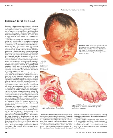
Plate 4-17                                                                                            Integumentary System

                                                                           CUTANEOUS MANIFESTATIONS OF LUPUS


       CUTANEOUS LUPUS (Continued)


       Treatment includes keeping the regions dry and warm
       by  avoiding  cold  exposure.  Patients  diagnosed  with
       pernio  probably  should  undergo  screening  for  lupus,
       because a small percentage of them actually have lupus
       chilblains.  Histological  evaluation  of  lupus  chilblains
       shows  a  dense  lymphocytic  infiltrate  with  some  areas
       of  thrombosis  of  small  vessels  and  a  lymphocytic
       vasculitis.
         The cutaneous findings seen in SLE are vast and can
       overlap with other forms of cutaneous lupus. Although
       the systemic findings are responsible for the morbidity
       and mortality, the cutaneous findings are often the pre-
       senting sign, and, if the clinician is aware, they can help                               Neonatal lupus. Neonatal lupus is transient
       make the diagnosis. The most important of the cutane-                                     in nature and is caused by maternal anti-
                                                                                                 bodies that cross the placenta. Newborns
       ous skin findings in SLE is the malar rash. This rash                                     are at risk for developing heart block.
       manifests as a tender, pink-to-red plaque or patch on                                     The cutaneous findings eventually resolve
       the cheeks and nose, mimicking the shape of a butterfly;                                  spontaneously.
       hence, it has been termed the “butterfly rash of lupus.”
       It  is  commonly  mistaken  for  rosacea,  and  vice  versa.
       Rosacea  typically  affects  a  wider  area  of  skin  and  is
       associated with more telangiectases and papulopustular
       lesions. The malar rash of lupus also spares the nasola-
       bial fold, which is an important clinical finding and a
       discriminating objective discovery. It is typically more
       prominent  during  systemic  flares  of  the  underlying
       SLE,  and  patients  can  appear  very  ill.  Patients  are
       exquisitely photosensitive, and the rash is exacerbated
       by exposure to ultraviolet light.
         Discoid lupus is also seen as a manifestation of sys-
       temic lupus, and it has the same clinical appearance as
       described  earlier.  Raynaud’s  phenomenon  is  well
       described, and a high percentage of patients with SLE
       report those symptoms. Alopecia was long used to help
       make the diagnosis of lupus. It is no longer part of the
       diagnostic criteria, but it can have significant psycho-
       logical  impact  on  the  patient.  Nail  and  capillary  nail
       fold changes are seen if looked for. The true incidence
       of these findings is unknown. Nail fold telangiectases
       and erythema are the two most common nail findings.
       Nail pitting, ridging, and alterations in the color of the
       lunula have also been reported. Lupus patients with nail
       changes have been found to have a higher incidence of
       mucosal ulcerations, which are another of the mucocu-
       taneous findings of SLE. Livedo reticularis is a fishnet-
       like pattern found typically on the lower extremities; it
       is  a  nonspecific  finding  but  has  been  reported  com-
       monly in lupus. It also occurs in many other skin and
       systemic diseases.                                                               Lupus chilblains. Tender red to purple macules
         Histology: The histological findings in all forms of                           and papules on the feet. Exacerbated by cold
       lupus  are  similar,  with  specific  forms  having  some   Lupus erythematosus disseminatus  and wet environments
       unique findings. Most forms show an interface derma-
       titis with hydropic changes in the basilar layer of the
       epidermis. A superficial and deep periadnexal lympho-
       cytic infiltrate is almost universally seen. Other connec-  Treatment: The treatment of cutaneous lupus is dif-  immediately, and patients should be screened routinely
       tive  tissue  diseases  (e.g.,  dermatomyositis)  can  have   ficult and must be tailored to the patient and the specific   by their family physician or rheumatologist for progres-
       similar  histological  findings.  Discoid  lupus  may  show   form of lupus. Potent topical corticosteroids may work   sion of the disease.
       scarring,  atrophy,  and  follicular  plugging  along  with   for a tiny lesion of discoid lupus, but they are not effec-  Specific  therapies  for  cutaneous  lupus  include  oral
       these  other  findings.  Lupus  panniculitis  is  unique  in   tive in lupus panniculitis. Universal treatment of cuta-  prednisone and hydroxychloroquine or chloroquine as
       that the inflammation is localized to the subcutaneous   neous lupus requires sun protection and sunscreen use.   the  typical  first-line  agents.  If  these  are  unsuccessful,
       tissue.  The  diagnosis  of  lupus  panniculitis  is  difficult   The  sunscreen  used  should  block  in  the  UVA  range,   quinacrine can be added. Other agents that have been
       and requires a host of special stains and clinical patho-  because this is the most active form of ultraviolet light   reported to be effective include dapsone, isotretinoin,
       logical correlation.                      that  exacerbates  lupus.  Smoking  should  be  ceased   and methotrexate.

       88                                                                                    THE NETTER COLLECTION OF MEDICAL ILLUSTRATIONS