
Page 106 - The Netter Collection of Medical Illustrations - Integumentary System_ Volume 4 ( PDFDrive )
P. 106
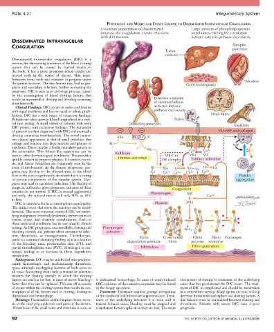
Plate 4-21                                                                                            Integumentary System

                                                      PHYSIOLOGY AND MOLECULAR EVENTS LEADING TO DISSEMINATED INTRAVASCULAR COAGULATION
                                                   Cutaneous presentation of disseminated        Large amounts of phospholipoprotein
                                                   intravascular coagulation. Livedo reticularis  membranes entering the circulation
                                                   with skin necrosis                            activate extrinsic pathway excessively.
       DISSEMINATED INTRAVASCULAR
       COAGULATION                                                                                                   Abruptio
                                                                                    Tumor                            placentae
                                                                                    necrosis
       Disseminated  intravascular  coagulation  (DIC)  is  a
       serious, life-threatening condition of the blood clotting
       system  that  can  be  caused  by  myriad  insults  to
       the  body.  It  has  a  grave  prognosis  unless  caught  and
       treated  early  in  the  course  of  disease.  Skin  mani-
       festations occur early and continue to progress unless                                                              Abortion
       the patient recovers. The skin lesions may lead to gan-                                Giant hemangioma
       grene and secondary infection, further worsening the
       prognosis. DIC is seen as an end-stage process, caused
       by  the  consumption  of  blood  clotting  factors,  that
       results in uncontrolled clotting and bleeding occurring                Extensive exposure
       simultaneously.                                                        of subendothelium
         Clinical Findings: DIC occurs in males and females                   activates intrinsic
       with equal incidence and has no racial or ethnic predi-                pathway excessively.
       lection.  DIC  has  a  wide  range  of  cutaneous  findings.                                       Head injury
       Patients are often gravely ill and hospitalized in a criti-                      Sepsis
       cal  care  setting.  A  small  subset  of  patients  with  early   Dissecting aneurysm                         Snakebite
       DIC present with cutaneous findings. The remainder
       of patients are first diagnosed with DIC and eventually                                                VIII:vWF and collagen
       develop cutaneous manifestations. The initial cutane-  XIIa  PK Kin XI
       ous clinical appearance is that of small petechiae that
       enlarge and coalesce into large macules and plaques of   XII                                                  ADP
       erythema. There may be a livedo reticularis pattern to             XIa
       the  extremities.  This  fishnet-like  appearance  can  be   Kallikrein                  Ca 2+  Ca 2+
       seen in other dermatological conditions. The petechiae                      VIII:C in    VIIa  VII
       quickly convert to purpuric plaques. Ulceration, necro-  Intrinsic activation  VIII complex  Extrinsic activation
       sis,  and  blister  formation  are  commonly  seen  in  the
       areas  of  involvement.  As  the  disease  progresses,  gan-         IX    IXa   X     Xa   Va  II  IIa
       grene may develop in the affected areas as the blood                 Ca    Ca    Ca 2+   Ca 2+    Ca 2+
                                                                                    2+
       flow to the skin is significantly decreased due to clotting                                                      Platelet
       of  various  components  of  the  vascular  system.  Gan-                                                       aggregation
       grene may lead to secondary infection. The finding of
       gangrene indicates a grave prognosis, and most of these                         Coagulation
       patients do not survive. If DIC is treated aggressively              Plasminogen       XIII
       and  early,  the  survival  rate  is  still  only  40%  to  50%                                  Ca 2+
       at best.
         DIC is considered to be a consumptive coagulopathy.                  Plasmin         XIIIa
       The initial event that starts the reaction can be multi-
       factorial. The most common causes of DIC are under-
       lying malignancy (especially leukemia), severe traumatic
       events,  sepsis,  and  obstetric  complications.  Each  of
       these associated conditions has its own specific clinical
       setting. As DIC progresses, uncontrollable clotting and   Plasminogen                 NH
       bleeding coexist, and patients often succumb to infec-  activator                        3
       tion,  thrombosis,  or  exsanguination.  Thrombocyto-           Fibrin     Cross-linked   Fibrin    Fibrin  Fibrinogen
       penia is a common laboratory finding, as is an elevation   degradation products  fibrin  polymer  monomer
       of  the  bleeding  time,  prothrombin  time  (PT),  and
       partial thromboplastin time (PTT). Fibrinogen is con-           Fibrinolysis              Fibrin generation
       sumed,  leading  to  an  increase  in  fibrin  degradation
       metabolites.
         Pathogenesis: DIC may be subdivided into predomi-
       nantly  hemorrhagic  and  predominantly  thrombotic
       types, although overlapping features of both occur in
       all cases. An inciting event such as trauma or infection
       initiates  the  clotting  cascade  in  which  the  clotting
       factors are used up (or lost, in cases of severe bleeding)   is  widespread  hemorrhage.  In  cases  of  sepsis-induced   component of therapy is treatment of the underlying
       faster than they can be replaced. This sets off a cascade   DIC, evidence of the causative organism may be found   cause that has precipitated the DIC event. The treat-
       of events within the clotting system that results in con-  in the biopsy specimen.  ment of DIC is complicated and should be undertaken
       sumption of all the factors used in clotting, leading to   Treatment: Treatment requires prompt recognition   in a critical care setting. Many agents are used to help
       thrombosis and hemorrhage.                of the condition and immediate supportive care. Treat-  decrease thrombosis and replace lost clotting factors. A
         Histology: Examination of skin biopsies shows necro-  ment  of  the  underlying  infection  is  a  must,  and  in   fine balance must be maintained between clotting and
       sis of the overlying epidermis and parts of the dermis.   trauma-induced  cases,  bleeding  must  be  stopped  and   thrombosis.  Patients  with  severe  DIC  have  a  poor
       Thrombosis of the small veins and arterioles is seen, as   coagulation factors replaced as they are lost. The main   prognosis.

       92                                                                                    THE NETTER COLLECTION OF MEDICAL ILLUSTRATIONS