
Page 107 - The Netter Collection of Medical Illustrations - Integumentary System_ Volume 4 ( PDFDrive )
P. 107
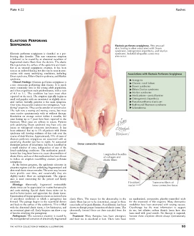
Plate 4-22                                                                                                           Rashes


        ELASTOSIS PERFORANS
        SERPIGINOSA                                                               Elastosis perforans serpiginosa. This unusual
                                                                                  skin finding is often associated with Down
                                                                                  syndrome, osteogenesis imperfecta, and Marfan
        Elastosis  perforans  serpiginosa  is  classified  as  a  per-            syndrome. Isolated idiopathic cases may
        forating  skin  disorder.  This  rare  cutaneous  eruption                also occur.
        is believed to be caused by an abnormal expulsion of
        fragmented elastic fibers from the dermis. The elastic
        fibers penetrate the surface of the epidermis and mani-
        fest  as  an  unusual  serpiginous  eruption.  It  has  been
        seen as an isolated finding but also can be seen in asso-
        ciation  with  many  underlying  conditions,  including                            Associations with Elastosis Perforans Serpiginosa
        Down syndrome, Ehlers-Danlos syndrome, and Marfan
        syndrome.                                                                             Acrogeria
          Clinical Findings: Elastosis perforans serpiginosa is                               Chronic renal failure
        a  rare  cutaneous  perforating  skin  disease.  It  is  much                         Down syndrome
        more  commonly  seen  in  the  young  adult  population,
        and it has a significant male predominance, with a ratio                              Ehlers-Danlos syndrome
        of  4 : 1  to  5 : 1.  The  condition  has  been  most  often                         Marfan syndrome
        reported on the neck. The eruption typically begins as                                Medications—penicillamine
        small red papules with an excoriated or slightly ulcer-                               Osteogenesis imperfecta
        ated  surface.  Initially,  pruritus  is  the  main  symptom.                         Pseudoxanthoma elasticum
        Over time, the papules coalesce into serpiginous, “wan-                               Rothmund-Thomson syndrome
        dering” eruptions. They can be annular or semicircular.                               Scleroderma
        The rash runs a waxing and waning course, but most
        cases  resolve  spontaneously  with  or  without  therapy.
        Resolution  on  average  occurs  within  6  months,  but
        cases lasting up to 5 years have been reported in the
        literature.  Most  cases  are  solitary  in  nature.  Patients
        with  underlying  Down  syndrome  may  have  only  one
        lesion  or  widespread  cutaneous  involvement.  It  has
        been estimated that up to 1% of patients with Down
        syndrome  will  develop  evidence  of  this  rash  over  the
        course of their lifetime. Approximately 33% of cases of
        elastosis  perforans  serpiginosa  are  associated  with  an
        underlying  disorder  (see  box  to  right).  An  autosomal
        dominant pattern of inheritance has been described in   Dense connective tissue
        a  small  number  of  cases,  independent  of  any  of  the
        listed underlying conditions. The medication penicil-
        lamine has long been known to cause abnormalities of              Longitudinal bundles
        elastic fibers, and use of this medication has been shown         of collagen and
        to  induce  an  eruption  resembling  elastosis  perforans        elastic fibers
        serpiginosa.
          As  the  lesions  progress,  the  epidermis  ulcerates  in
        pinpoint regions and the underlying fragmentized and
        abnormal elastic tissue extrudes. The areas may become
        more  pruritic  over  time,  and  occasionally  they  are
        slightly  tender.  Most  are  asymptomatic.  The  appear-
        ance  is  most  concerning  for  the  patient  and  family
        members.                                                                           Fibroblast       Transverse fibers of
          Histology:  Abnormally  fragmented  eosinophilic                                 nuclei           loose connective tissue
        elastic tissue can be appreciated on routine hematoxylin
        and eosin staining. Special elastic tissue stains can be
        used to better isolate and appreciate the elastic tissue.
        Examination of biopsy specimens shows an isolated area
        of  acanthotic  epidermis  in  which  a  passageway  has   elastic  fibers.  The  reason  for  the  abnormality  in  the   no  randomized,  prospective,  placebo-controlled  trials
        formed.  The  passage  begins  in  the  superficial  dermis   elastic fibers has yet to be determined, except in those   for  the  treatment  of  this  eruption.  Many  destructive
        and leads to the surface of the epidermis. This is filled   cases induced by penicillamine. Penicillamine has been   modalities  have  been  attempted  with  varying  success.
        with the abnormal elastic tissue, a few histiocytes, and   shown to disrupt proper formation of elastic tissue. The   Cryotherapy  has  the  most  information  to  support
        an occasional giant cell. Early biopsies can show a cap   abnormally formed fibers are then extruded from the   its  use,  but  ablative  carbon  dioxide  lasers  have  also
        of keratin overlying the passageway.      dermis.                                   been used with good results. No therapy is required,
          Pathogenesis: The cutaneous eruption is caused by   Treatment:  Many  therapies  have  been  attempted,   because  these  eruptions  almost  always  spontaneously
        the transepidermal extrusion of abnormally fragmented   and  their  use  is  anecdotal  at  best.  There  have  been     remit.


        THE NETTER COLLECTION OF MEDICAL ILLUSTRATIONS                                                                           93