
Page 111 - The Netter Collection of Medical Illustrations - Integumentary System_ Volume 4 ( PDFDrive )
P. 111
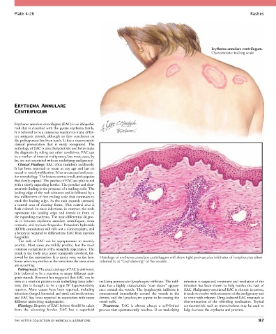
Plate 4-26                                                                                                           Rashes


                                                                                                          Erythema annulare centrifugum.
                                                                                                          Characteristic trailing scale


        ERYTHEMA ANNULARE
        CENTRIFUGUM


        Erythema annulare centrifugum (EAC) is an idiopathic
        rash that is classified with the gyrate erythema family.
        It is believed to be a cutaneous reaction to many differ-
        ent antigenic stimuli, although no firm conclusion on
        the pathogenesis has been made. It has a characteristic
        clinical  presentation  that  is  easily  recognized.  The
        pathology of EAC is also characteristic and helps make
        the diagnosis by ruling out other conditions. EAC can
        be a marker of internal malignancy, but most cases, by
        far, are not associated with an underlying malignancy.
          Clinical Findings: EAC often manifests insidiously.
        It  has  been  reported  to  occur  at  any  age  and  has  no
        sexual or racial predilection. It has an unusual and pecu-
        liar morphology. The lesions start as small, pink papules
        that slowly expand. The patches of EAC are pink to red
        with a slowly expanding border. The peculiar and char-
        acteristic finding is the presence of a trailing scale. The
        leading edge of the rash advances and is followed by a
        few millimeters of fine trailing scale that continues to
        track  the  leading  edge.  As  the  rash  expands  outward,
        a  central  area  of  clearing  forms.  This  central  area  is
        flesh colored. In tinea infections, in contrast, the scale
        represents  the  leading  edge  and  travels  in  front  of
        the expanding erythema. The main differential diagno-
        sis  is  between  erythema  annulare  centrifugum,  tinea
        corporis, and mycosis fungoides. Potassium hydroxide
        (KOH) examination will rule out a dermatophyte, and
        a biopsy is required to differentiate EAC from mycosis
        fungoides.
          The rash of EAC can be asymptomatic to severely
        pruritic.  Most  cases  are  mildly  pruritic,  but  the  most
        common complaint is of the unsightly appearance. The
        trunk is the body area most commonly involved, fol-
        lowed by the extremities. It is rarely seen on the face.   Histology of erythema annulare centrifugum will show tight perivascular infiltrates of lymphocytes often
        Some areas may resolve at the same time that new areas   referred to as “coat sleeving” of the vessels.
        are occurring.
          Pathogenesis: The exact etiology of EAC is unknown.
        It is believed to be a reaction to many different anti-
        genic stimuli. Research has suggested that EAC can be
        seen as a reaction pattern to an underlying tinea infec-  and deep perivascular lymphocytic infiltrate. The infil-  infection is suspected, treatment and resolution of the
        tion;  this  is  thought  to  be  a  type  IV  hypersensitivity   trate  has  a  highly  characteristic  “coat  sleeve”  appear-  infection  has  been  shown  to  help  resolve  the  rash  of
        reaction.  Many  causes  have  been  reported,  including   ance around the vessels. The lymphocytic infiltrate is   EAC. Malignancy-associated EAC is chronic in nature;
        infections (fungal, bacterial, and viral) and medications,   concentrated  immediately  around  the  vessels  in  the   it tends to resolve with treatment of the malignancy and
        and EAC has been reported in association with many   dermis, and the lymphocytes appear to be coating the   to recur with relapses. Drug-induced EAC responds to
        different underlying malignancies.        vessel walls.                             discontinuation  of  the  offending  medication.  Topical
          Histology: Biopsies of EAC lesions should be taken   Treatment:  EAC  is  almost  always  a  self-limited   corticosteroids  such  as  triamcinolone  may  be  used  to
        from  the  advancing  border.  EAC  has  a  superficial     process  that  spontaneously  resolves.  If  an  underlying   help decrease the erythema and pruritus.


        THE NETTER COLLECTION OF MEDICAL ILLUSTRATIONS                                                                           97