
Page 112 - The Netter Collection of Medical Illustrations - Integumentary System_ Volume 4 ( PDFDrive )
P. 112
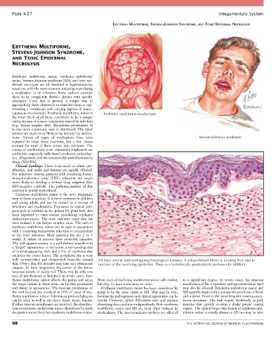
Plate 4-27                                                                                            Integumentary System

                                                        ERYTHEMA MULTIFORME, STEVENS-JOHNSON SYNDROME, AND TOXIC EPIDERMAL NECROLYSIS


       ERYTHEMA MULTIFORME,
       STEVENS-JOHNSON SYNDROME,
       AND TOXIC EPIDERMAL
       NECROLYSIS


       Erythema  multiforme  minor,  erythema  multiforme
       major, Stevens-Johnson syndrome (SJS), and toxic epi-
       dermal  necrolysis  are  all  classified  as  hypersensitivity
       reactions, with the most common initiating event being
       a  medication  or  an  infection.  Some  authors  consider
       these  to  be  completely  distinct  entities  with  specific
       etiologies.  Until  that  is  proven,  a  simple  way  of
       approaching these diseases is to consider them as rep-
       resenting a continuum with varying degrees of muco-
       cutaneous involvement. Erythema multiforme minor is   Erythema multiforme exudativum
       the most likely of all these conditions to be a unique
       entity, because it is more commonly caused by infection
       (e.g., herpes simplex virus, Mycoplasma pneumoniae). It
       is also more commonly seen in childhood. The other
       entities are much more likely to be initiated by medica-
       tions.  Almost  all  types  of  medications  have  been                                     Stevens-Johnson syndrome
       reported  to  cause  these  reactions,  but  a  few  classes
       account  for  most  of  these  severe  skin  reactions.  The
       classes of medications most commonly implicated are
       antibiotics (especially sulfa-based products), antiepilep-
       tics, allopurinol, and the nonsteroidal antiinflammatory
       drugs (NSAIDs).
         Clinical Findings: There is no racial or ethnic pre-
       dilection, and males and females are equally affected.
       For unknown reasons, patients with coexisting human
       immunodeficiency  virus  (HIV)  infection  are  much
       more  likely  to  develop  a  serious  drug  eruption  than
       HIV-negative  controls.  The  pathomechanism  of  this
       reaction is poorly understood.
         Erythema multiforme minor is the most frequently
       seen of these eruptions. It is more common in children
       and  young  adults  and  can  be  caused  by  a  myriad  of
       infections and medications. Exposures to topical anti-
       gens such as urushiol in the poison ivy plant have also
       been  reported  to  cause  rashes  resembling  erythema
       multiforme minor. The most common cause that has
       been isolated is the herpes simplex virus. The rash of
       erythema multiforme minor can be seen in association
       with a coexisting herpesvirus infection or independent
       of  the  viral  infection.  Most  episodes  last  for  2  to  3
       weeks.  A  subset  of  patients  have  recurrent  episodes.
       The rash appears acutely as a well-defined macule with
       a “target” appearance—a red center, a surrounding area
       of normal-appearing skin, and a rim of erythema that
       encircles the entire lesion. The peripheral rim is very
       well  circumscribed  and  demarcated  from  the  normal   All have similar and overlapping histological features. A subepidermal blister is forming here due to
       skin. Over a day, the macules may turn into edematous   necrosis of the overlying epidermis. There is a lymphocytic predominate perivascular infiltrate.
       plaques.  As  time  progresses,  the  center  of  the  lesion
       becomes purple or dusky red. There may be only one
       area of involvement or hundreds in severe cases. Ery-
       thema  multiforme  minor  affects  the  palms  and  soles;   Most cases of erythema multiforme minor self-resolve,   to  a  significant  degree.  In  severe  cases,  the  mucosal
       the target lesions in these areas can be very prominent   but they do have a tendency to recur.  membranes of the respiratory and gastrointestinal tract
       and classic in appearance. The mucous membranes of   Erythema multiforme major has been considered by   may also be affected. Erythema multiforme major and
       the oral mucosa are involved in 20% of cases of ery-  many to be the same entity as SJS. This may be true,   SJS typically begin with a nonspecific prodrome of fever
       thema multiforme minor. Edematous pink-red plaques   because the pathogenesis and clinical appearance can be   and malaise. Fever is the most frequent nonmucocuta-
       can be seen, as well as the more classic target lesions.   similar. However, subtle differences exist and warrant   neous  symptom.  The  rash  begins  insidiously  as  pink
       If other mucous membranes are involved, the classifica-  classifying this condition independently. Both erythema   macules  that  quickly  develop  a  dusky  purple  central
       tion of erythema multiforme minor should not be used;   multiforme major and SJS are most often induced by   region. The typical target-like lesion of erythema mul-
       the patient more likely has erythema multiforme major.   medications. The mucocutaneous surfaces are affected   tiforme minor is usually absent in SJS but may be seen

       98                                                                                    THE NETTER COLLECTION OF MEDICAL ILLUSTRATIONS