
Page 114 - The Netter Collection of Medical Illustrations - Integumentary System_ Volume 4 ( PDFDrive )
P. 114
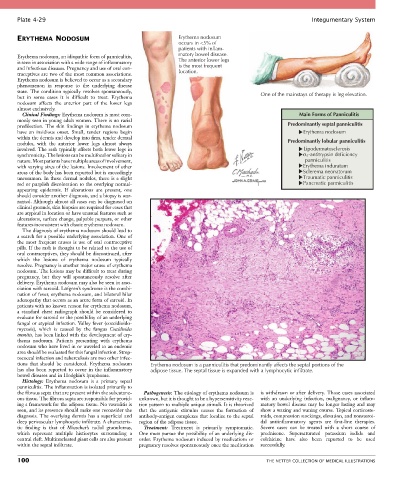
Plate 4-29                                                                                            Integumentary System


       ERYTHEMA NODOSUM                                        Erythema nodosum
                                                               occurs in <5% of
                                                               patients with inflam-
       Erythema nodosum, an idiopathic form of panniculitis,   matory bowel disease.
       is seen in association with a wide range of inflammatory   The anterior lower legs
       and infectious diseases. Pregnancy and use of oral con-  is the most frequent
       traceptives are two of the most common associations.    location.
       Erythema nodosum is believed to occur as a secondary
       phenomenon  in  response  to  the  underlying  disease
       state.  The  condition  typically  resolves  spontaneously,                         One of the mainstays of therapy is leg elevation.
       but  in  some  cases  it  is  difficult  to  treat.  Erythema
       nodosum  affects  the  anterior  part  of  the  lower  legs
       almost exclusively.
         Clinical Findings: Erythema nodosum is most com-                                               Main Forms of Panniculitis
       monly seen in young adult women. There is no racial
       predilection.  The  skin  findings  in  erythema  nodosum                                    Predominantly septal panniculitis
       have  an  insidious  onset.  Small,  tender  regions  begin                                        Erythema nodosum
       within the dermis and develop into firm, tender dermal
       nodules,  with  the  anterior  lower  legs  almost  always                                   Predominantly lobular panniculitis
       involved. The rash typically affects both lower legs in                                             Lipodermatosclerosis
       synchronicity. The lesions can be multifocal or solitary in                                         α 1-antitrypsin deficiency
       nature. Most patients have multiple areas of involvement,                                           panniculitis
       with varying sizes of the lesions. Involvement of other                                             Erythema induratum
       areas of the body has been reported but is exceedingly                                              Sclerema neonatorum
       uncommon. In these dermal nodules, there is a slight                                                Traumatic panniculitis
       red or purplish discoloration to the overlying normal-                                              Pancreatic panniculitis
       appearing  epidermis.  If  ulcerations  are  present,  one
       should consider another diagnosis, and a biopsy is war-
       ranted. Although almost all cases can be diagnosed on
       clinical grounds, skin biopsies are required for cases that
       are atypical in location or have unusual features such as
       ulcerations, surface change, palpable purpura, or other
       features inconsistent with classic erythema nodosum.
         The diagnosis of erythema nodosum should lead to
       a search for a possible underlying association. One of
       the  most  frequent  causes  is  use  of  oral  contraceptive
       pills. If the rash is thought to be related to the use of
       oral contraceptives, they should be discontinued, after
       which  the  lesions  of  erythema  nodosum  typically
       resolve. Pregnancy is another major cause of erythema
       nodosum. The lesions may be difficult to treat during
       pregnancy,  but  they  will  spontaneously  resolve  after
       delivery. Erythema nodosum may also be seen in asso-
       ciation with sarcoid. Löfgren’s syndrome is the combi-
       nation of fever, erythema nodosum, and bilateral hilar
       adenopathy that occurs as an acute form of sarcoid. In
       patients with no known reason for erythema nodosum,
       a  standard  chest  radiograph  should  be  considered  to
       evaluate for sarcoid or the possibility of an underlying
       fungal or atypical infection. Valley fever (coccidioido-
       mycosis),  which  is  caused  by  the  fungus  Coccidioides
       immitis, has been linked with the development of ery-
       thema  nodosum.  Patients  presenting  with  erythema
       nodosum who have lived in or traveled to an endemic
       area should be evaluated for this fungal infection. Strep-
       tococcal infection and tuberculosis are two other infec-
       tions  that  should  be  considered.  Erythema  nodosum   Erythema nodosum is a panniculitis that predominantly affects the septal portions of the
       has  also  been  reported  to  occur  in  the  inflammatory   adipose tissue. The septal tissue is expanded with a lymphocytic infiltrate.
       bowel diseases and in Hodgkin’s lymphoma.
         Histology:  Erythema  nodosum  is  a  primary  septal
       panniculitis. The inflammation is isolated primarily to
       the fibrous septa that are present within the subcutane-  Pathogenesis: The etiology of erythema nodosum is   is withdrawn or after delivery. Those cases associated
       ous tissue. The fibrous septa are responsible for provid-  unknown, but it is thought to be a hypersensitivity reac-  with  an  underlying  infection,  malignancy,  or  inflam-
       ing a framework for the adipose tissue. No vasculitis is   tion pattern to multiple unique stimuli. It is theorized   matory bowel disease may be longer lasting and may
       seen, and its presence should make one reconsider the   that  the  antigenic  stimulus  causes  the  formation  of   show a waxing and waning course. Topical corticoste-
       diagnosis. The overlying dermis has a superficial and   antibody-antigen complexes that localize to the septal   roids, compression stockings, elevation, and nonsteroi-
       deep perivascular lymphocytic infiltrate. A characteris-  region of the adipose tissue.  dal  antiinflammatory  agents  are  first-line  therapies.
       tic  finding  is  that  of  Miescher’s  radial  granulomas,   Treatment:  Treatment  is  primarily  symptomatic.   Severe  cases  can  be  treated  with  a  short  course  of
       which  represent  multiple  histiocytes  surrounding  a   One must pursue the possibility of an underlying dis-  prednisone.  Supersaturated  potassium  iodide  and
       central cleft. Multinucleated giant cells are also present   order. Erythema nodosum induced by medications or   colchicine  have  also  been  reported  to  be  used
       within the septal infiltrate.             pregnancy resolves spontaneously once the medication   successfully.

       100                                                                                   THE NETTER COLLECTION OF MEDICAL ILLUSTRATIONS