
Page 113 - The Netter Collection of Medical Illustrations - Integumentary System_ Volume 4 ( PDFDrive )
P. 113
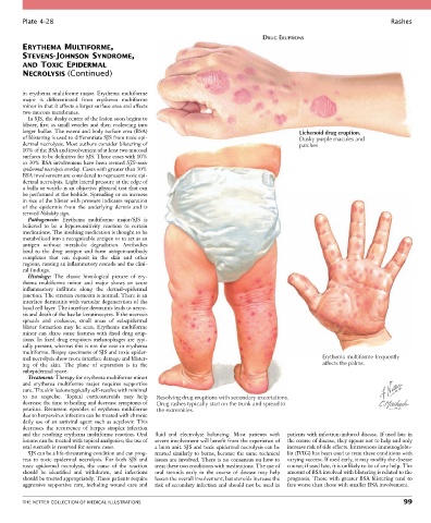
Plate 4-28                                                                                                           Rashes

                                                                                    DRUG ERUPTIONS
        ERYTHEMA MULTIFORME,
        STEVENS-JOHNSON SYNDROME,
        AND TOXIC EPIDERMAL
        NECROLYSIS (Continued)


        in  erythema  multiforme  major.  Erythema  multiforme
        major  is  differentiated  from  erythema  multiforme
        minor in that it affects a larger surface area and affects
        two mucous membranes.
          In SJS, the dusky center of the lesion soon begins to
        blister, first as small vesicles and then coalescing into
        larger bullae. The extent and body surface area (BSA)                                   Lichenoid drug eruption.
        of blistering is used to differentiate SJS from toxic epi-                              Dusky purple macules and
        dermal necrolysis. Most authors consider blistering of                                  patches
        10% of the BSA and involvement of at least two mucosal
        surfaces to be definitive for SJS. Those cases with 10%
        to 30% BSA involvement have been termed SJS–toxic
        epidermal necrolysis overlap. Cases with greater than 30%
        BSA involvement are considered to represent toxic epi-
        dermal necrolysis. Light lateral pressure at the edge of
        a bulla or vesicle is an objective physical test that can
        be performed at the bedside. Spreading or an increase
        in size of the blister with pressure indicates separation
        of  the  epidermis  from  the  underlying  dermis  and  is
        termed Nikolsky sign.
          Pathogenesis:  Erythema  multiforme  major/SJS  is
        believed  to  be  a  hypersensitivity  reaction  to  certain
        medications. The insulting medication is thought to be
        metabolized into a recognizable antigen or to act as an
        antigen  without  metabolic  degradation.  Antibodies
        bind  to  the  drug  antigen  and  form  antigen-antibody
        complexes  that  can  deposit  in  the  skin  and  other
        regions, causing an inflammatory cascade and the clini-
        cal findings.
          Histology:  The  classic  histological  picture  of  ery-
        thema  multiforme  minor  and  major  shows  an  acute
        inflammatory  infiltrate  along  the  dermal-epidermal
        junction. The stratum corneum is normal. There is an
        interface dermatitis with vacuolar degeneration of the
        basal cell layer. The interface dermatitis leads to necro-
        sis and death of the basilar keratinocytes. If the necrosis
        spreads  and  coalesces,  small  areas  of  subepidermal
        blister  formation  may  be  seen.  Erythema  multiforme
        minor  can  share  some  features  with  fixed  drug  erup-
        tions. In fixed drug eruptions melanophages are typi-
        cally present, whereas this is not the case in erythema
        multiforme. Biopsy specimens of SJS and toxic epider-
        mal necrolysis show more interface damage and blister-                                         Erythema multiforme frequently
        ing  of  the  skin.  The  plane  of  separation  is  in  the                                   affects the palms.
        subepidermal space.
          Treatment: Therapy for erythema multiforme minor
        and  erythema  multiforme  major  requires  supportive
        care. The skin lesions typically self-resolve with minimal
        to  no  sequelae.  Topical  corticosteroids  may  help   Resolving drug eruptions with secondary excoriations.
        decrease the time to healing and decrease symptoms of   Drug rashes typically start on the trunk and spread to
        pruritus.  Recurrent  episodes  of  erythema  multiforme   the extremities.
        due to herpesvirus infection can be treated with chronic
        daily use of an antiviral agent such as acyclovir. This
        decreases  the  recurrence  of  herpes  simplex  infection
        and the resulting erythema multiforme reaction. Oral   fluid  and  electrolyte  balancing.  Most  patients  with   patients with infection-induced disease. If used late in
        lesions can be treated with topical analgesics; the use of   severe involvement will benefit from the experience of   the course of disease, they appear not to help and only
        oral steroids is reserved for severe cases.  a burn unit. SJS and toxic epidermal necrolysis can be   increase risk of side effects. Intravenous immunoglobu-
          SJS can be a life-threatening condition and can prog-  treated similarly to burns, because the same technical   lin (IVIG) has been used to treat these conditions with
        ress  to  toxic  epidermal  necrolysis.  For  both  SJS  and   issues are involved. There is no consensus on how to   varying success. If used early, it may modify the disease
        toxic  epidermal  necrolysis,  the  cause  of  the  reaction   treat these two conditions with medications. The use of   course; if used late, it is unlikely to be of any help. The
        should  be  identified  and  withdrawn,  and  infections   oral  steroids  early  in  the  course  of  disease  may  help   amount of BSA involved with blistering is related to the
        should be treated appropriately. These patients require   lessen the overall involvement, but steroids increase the   prognosis. Those with greater BSA blistering tend to
        aggressive supportive care, including wound care and   risk of secondary infection and should not be used in   fare worse than those with smaller BSA involvement.


        THE NETTER COLLECTION OF MEDICAL ILLUSTRATIONS                                                                           99