
Page 147 - The Netter Collection of Medical Illustrations - Integumentary System_ Volume 4 ( PDFDrive )
P. 147
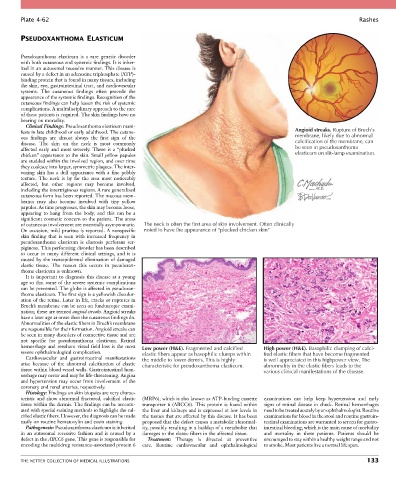
Plate 4-62                                                                                                           Rashes


        PSEUDOXANTHOMA ELASTICUM


        Pseudoxanthoma  elasticum  is  a  rare  genetic  disorder
        with both cutaneous and systemic findings. It is inher-
        ited in an autosomal recessive manner. This disease is
        caused by a defect in an adenosine triphosphate (ATP)-
        binding protein that is found in many tissues, including
        the skin, eye, gastrointestinal tract, and cardiovascular
        systems.  The  cutaneous  findings  often  precede  the
        appearance of the systemic findings. Recognition of the
        cutaneous findings can help lessen the risk of systemic
        complications. A multidisciplinary approach to the care
        of these patients is required. The skin findings have no
        bearing on mortality.
          Clinical Findings: Pseudoxanthoma elasticum mani-
        fests in late childhood or early adulthood. The cutane-                                        Angioid streaks. Rupture of Bruch’s
        ous  findings  are  almost  always  the  first  sign  of  the                                  membrane, likely due to abnormal
        disease.  The  skin  on  the  neck  is  most  commonly                                         calcification of the membrane, can
        affected  early  and  most  severely.  There  is  a  “plucked                                  be seen in pseudoxanthoma
        chicken” appearance to the skin. Small yellow papules                                          elasticum on slit-lamp examination.
        are studded within the involved region, and over time
        they coalesce into larger, symmetric plaques. The inter-
        vening  skin  has  a  dull  appearance  with  a  fine  pebbly
        texture.  The  neck  is  by  far  the  area  most  noticeably
        affected,  but  other  regions  may  become  involved,
        including the intertriginous regions. A rare generalized
        cutaneous form has been reported. The mucous mem-
        branes  may  also  become  involved  with  tiny  yellow
        papules. As time progresses, the skin may become loose,
        appearing  to  hang  from  the  body,  and  this  can  be  a
        significant cosmetic concern to the patient. The areas
        of cutaneous involvement are essentially asymptomatic.   The neck is often the first area of skin involvement. Often clinically
        On occasion, mild pruritus is reported. A nonspecific   noted to have the appearance of “plucked chicken skin”
        skin  finding  that  is  seen  with  increased  frequency  in
        pseudoxanthoma  elasticum  is  elastosis  perforans  ser-
        piginosa. This perforating disorder has been described
        to  occur  in  many  different  clinical  settings,  and  it  is
        caused by the transepidermal elimination of damaged
        elastic  tissue.  The  reason  this  occurs  in  pseudoxan-
        thoma elasticum is unknown.
          It is important to diagnosis this disease at a young
        age so that some of the severe systemic complications
        can be prevented. The globe is affected in pseudoxan-
        thoma elasticum. The first sign is a yellowish discolor-
        ation of the retina. Later in life, cracks or ruptures in
        Bruch’s membrane can be seen on funduscopic exami-
        nation; these are termed angioid streaks. Angioid streaks
        have a later age at onset than the cutaneous findings do.
        Abnormalities of the elastic fibers in Bruch’s membrane
        are responsible for their formation. Angioid streaks can
        be seen in many disorders of connective tissue and are
        not  specific  for  pseudoxanthoma  elasticum.  Retinal
        hemorrhage and resultant visual field loss is the most   Low power (H&E). Fragmented and calcified  High power (H&E). Basophilic clumping of calci-
        severe ophthalmological complication.     elastic fibers appear as basophilic clumps within  fied elastic fibers that have become fragmented
          Cardiovascular  and  gastrointestinal  manifestations   the middle to lower dermis. This is highly  is well appreciated in this highpower view. The
        arise  because  of  the  abnormal  calcification  of  elastic   characteristic for pseudoxanthoma elasticum.  abnormality in the elastic fibers leads to the
        tissue within blood vessel walls. Gastrointestinal hem-                             various clinical manifestations of the disease.
        orrhage may occur and may be life-threatening. Angina
        and hypertension may occur from involvement of the
        coronary and renal arteries, respectively.
          Histology: Findings on skin biopsies are very charac-
        teristic  and  show  abnormal  fractured,  calcified  elastic   (MRP6), which is also known as ATP-binding cassette   examinations  can  help  keep  hypertension  and  early
        tissue within the dermis. The findings can be accentu-  transporter  6  (ABCC6).  This  protein  is  found  within   signs of retinal disease in check. Retinal hemorrhages
        ated with special staining methods to highlight the cal-  the liver and kidneys and is expressed at low levels in   need to be treated acutely by an ophthalmologist. Routine
        cified elastic fibers. However, the diagnosis can be made   the tissues that are affected by this disease. It has been   examinations for blood in the stool and routine gastroin-
        easily on routine hematoxylin and eosin staining.  proposed that the defect causes a metabolic abnormal-  testinal examinations are warranted to screen for gastro-
          Pathogenesis: Pseudoxanthoma elasticum is inherited   ity, possibly resulting in a buildup of a metabolite that   intestinal bleeding, which is the main cause of morbidity
        in  an  autosomal  recessive  fashion  and  is  caused  by  a   damages to the elastic fibers in the affected tissue.  and  mortality  in  these  patients.  Patients  should  be
        defect in the ABCC6 gene. This gene is responsible for   Treatment:  Therapy  is  directed  at  preventive   encouraged to stay within a healthy weight range and not
        encoding the multidrug resistance–associated protein 6   care.  Routine  cardiovascular  and  ophthalmological   to smoke. Most patients live a normal life span.


        THE NETTER COLLECTION OF MEDICAL ILLUSTRATIONS                                                                          133