
Page 150 - The Netter Collection of Medical Illustrations - Integumentary System_ Volume 4 ( PDFDrive )
P. 150
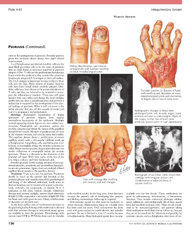
Plate 4-65                                                                                            Integumentary System

                                                                                  PSORIATIC ARTHRITIS


       PSORIASIS (Continued)


       clues to the pathogenesis of psoriasis. Psoriatic patients
       given  this  medicine  almost  always  have  rapid  clinical
       improvement.
         T-cell lymphocytes and dermal dendritic cells are the
       most likely precursor cells to be the cause of psoriasis;   Pitting, discoloration, and erosion
       they are both found in increased numbers in psoriatic   of fingernails with fusiform swelling
       plaques. CD8+ T cells are the predominant lymphocyte   of distal interphalangeal joints
       found within the epidermis; they contain the cutaneous
       lymphocyte antigen (CLA) antigen on their cell surface.
       The CLA antigen is important because it directs these
       cells  into  the  skin.  Many  subsets  of  dermal  dendritic
       cells  have  been  found  within  psoriatic  plaques.  Den-
       dritic cells have been shown to be potent stimulators of                                      Psoriatic patches on dorsum of hand
       T cells, and they are believed to be required to propa-                                       with swelling and distortion of many
       gate  the  inflammatory  reaction.  These  two  cell  types                                   interphalangeal joints and shortening
       interact with each other and change the local cytokine                                        of fingers due to loss of bone mass
       profile into one that is proinflammatory and provides a
       milieu that is required for the development of the clini-
       cal findings of psoriasis. What is still unknown is the
       initial stimulus that sets off this cascade of events and
       how it is propagated and perpetuated.                                                     Radiographic changes in distal inter-
         Histology:  Histological  examination  of  biopsy                                       phalangeal joint. Left, In early stages, bone
       specimens  of  psoriasis  vulgaris  show  regular                                         erosions are seen at joint margins. Right, In
       psoriasiform  hyperplasia  of  the  epidermis.  Multiple                                  late stages, further loss of bone mass
       normal-appearing mitotic figures are seen within kera-                                    produces “pencil point in cup” appearance.
       tinocytes.  Neutrophils  are  prominent  within  the
       stratum corneum and within the lumen of the papillary
       dermal blood vessels. Mounds of parakeratosis are seen
       in the stratum corneum and contain many neutrophils.
       The  papillary  dermis  shows  a  proliferation  of  ectatic
       capillary vessels with a perivascular infiltrate made up
       of lymphocytes, Langerhans cells, and histiocytes. Col-
       lections of neutrophils within the stratum corneum are
       called Munro microabscesses. Kogoj microabscesses are
       similar  collections  of  neutrophils  within  the  stratum
       spinosum. There is a decrease in the thickness of the
       granular cell layer. With time, some of the tips of the
       rete ridges coalesce and form thickened ends.
         Pustular psoriasis shows varying amounts of intraepi-
       dermal pustules; acanthosis and psoriasiform hyperpla-
       sia are not prominent. Again, there are multiple dilated
       capillary blood vessels in the papillary dermis.
         Treatment: There is no cure for psoriasis. Treatment                                    Radiograph of sacroiliac joints shows thin
       should  be  based  on  the  amount  and  location  of  the                                cartilage with irregular surface and
       psoriatic plaques and consideration of the psychological   Toes with sausage-like swelling,  condensation of adjacent bone in
       well-being  of  the  affected  individual.  Small  areas  in   skin lesions, and nail changes  sacrum and ilia.
       discrete locations can be treated with topical corticoste-
       roids,  anthralin,  tar  compounds,  or  vitamin  D  or  A
       analogues  or  left  alone  without  therapy.  Ultraviolet
       therapy with natural sunlight, narrow-band ultraviolet   with excellent results. In the long term, these therapies   available  over  the  last  decade.  These  medications  are
       B light (UVB), or psoralen + ultraviolet A light (PUVA)   increase  the  patient’s  risk  of  developing  skin  cancers,   given  by  subcutaneous,  intramuscular,  or  intravenous
       has been used with great success. Often, combinations   and lifelong dermatologic follow-up is required.  injection.  They  include  etanercept,  alefacept,  adalim-
       of therapies are implemented.               Oral systemic agents are also used for moderate to   umab, infliximab, and ustekinumab. All of these agents
         As the body surface area of involvement increases or   severe psoriasis. Methotrexate taken on a weekly basis   have had excellent response rates. They are all consid-
       the psychological well-being of the individual is affected   has  been  used  for  years.  Oral  cyclosporine  has  been   ered  to  be  immunosuppressive,  and  patients  taking
       such that systemic therapy is warranted, many agents   used with great success for erythrodermic and pustular   these medications need close clinical follow-up, because
       are available to treat the psoriasis. Phototherapy with   psoriasis. Its use is limited to 6 to 12 months because   they are at increased risk for infections and possibly for
       narrow-band UVB or PUVA has been used for decades   of nephrotoxicity. Many biological agents have become   systemic cancers, such as lymphoma, after years of use.

       136                                                                                   THE NETTER COLLECTION OF MEDICAL ILLUSTRATIONS