
Page 186 - The Netter Collection of Medical Illustrations - Integumentary System_ Volume 4 ( PDFDrive )
P. 186
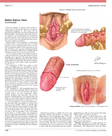
Plate 6-11                                                                                            Integumentary System

                                                                          LESIONS OF HERPES SIMPLEX (CONTINUED)


       HERPES SIMPLEX VIRUS
       (Continued)


         Recurrent  episodes  of  genital  herpes  produce  a
       milder version of the primary infection. The systemic                            Ulcerative lesions of genitalia
       constitutional  symptoms  are  often  absent,  but  the                          seen in chronic HSV and during
       grouped vesicles and ulcers can cause excruciating pain                          healing of vesicular stage
       and social stigma. The frequency and severity of recur-
       rent episodes in an individual patient are variable and
       impossible to predict. A generalization can be made that
       those who have more severe primary infections tend to
       have more relentless recurrences.
         Herpetic whitlow is the name given to a specific form
       of  infection  that  is  most  commonly  seen  in  medical
       laboratory workers and health care providers. It occurs
       from accidental inoculation of the herpesvirus into the
       skin. The finger is the area most commonly involved,
       because of accidental needle sticks. A painful primary
       viral infection may occur at the site of inoculation.
         Eczema herpeticum, Kaposi’s varicelliform eruption,
       is often encountered in a young child with severe atopic
       dermatitis who is exposed to the herpesvirus. Because
       of the widespread skin disease, the virus is able to infect
       a large surface area of the body. This results in extensive
       skin involvement with multiple vesicles and punched-
       out ulcerations.
         The  transmission  of  HSV  from  mother  to  child                      Herpes progenitalis
       during  the  birthing  process  is  of  significant  concern,
       and  mothers  with  active  HSV  disease  at  the  time  of
       delivery most likely should undergo cesarean section to                                Tender grouped vesicles on a red base
       help decrease the risk of transmission. Neonatal HSV
       infection is a life-threatening disease. The neonate may
       have widespread multiorgan disease, with CNS involve-
       ment being the major cause of morbidity and mortality.                  Vesicles
       Temporal  lobe  involvement  can  lead  to  seizures,
       encephalitis, and death. The skin is always infected, and
       this is a clue for the clinician to search for other organ
       system involvement, especially involvement of the CNS
       and the eye. Ocular infection can lead to severe corneal
       scarring and blindness.                                       Ruptured vesicle
         HSV  encephalitis  is  a  life-threatening  disease  that   causing a thin
       causes a necrotizing encephalitis. Patients complain of       erosion
       an  acute  onset  of  fever  and  headache,  with  rapidly
       evolving  seizures  and  focal  neurological  deficits.
       Without  treatment,  coma  and  death  occur  in  three
       quarters of affected patients. The temporal lobes and
       insula are almost always affected. Prompt recognition
       and therapy have decreased the mortality rate to 1 in 4.
         A Tzanck preparation is a long-used bedside proce-
       dure that takes only a few minutes to perform and is
       positive  in  cases  of  HSV1,  HSV2,  or  varicella-zoster
       virus (VZV) infection. The procedure does not differ-                          Herpes genitalis. Regional adenoopathy is often appreciated.
       entiate among the three viruses. However, HSV infec-
       tion can be distinguished from varicella clinically. The
       procedure is done by unroofing a vesicle and scraping
       its base with a no. 15 blade scalpel. The scrapings are   Rapid immunostaining is available and can be used   sample fluoresces with one of the specific viral stains.
       placed on a glass slide and allowed to air dry for 1 to 2   with  high  sensitivity  and  specificity  to  diagnose  and   This test takes 1 to 2 hours to perform.
       minutes. A blue stain such as Giemsa or toluidine blue   differentiate the various herpesvirus types. This form   Viral  tissue  cultures  can  also  be  performed  to  dif-
       is applied for 60 seconds and then gently rinsed off. The   of direct fluorescent antibody (DFA) testing is similar   ferentiate the HSV types, but the results can take days
       slide is dried, mineral oil is applied, and the preparation   to the Tzanck preparation. As in the Tzanck prepara-  to  1  week  to  obtain.  This  is  the  most  sensitive  and
       is covered with a microscope cover slip. It is then ready   tion, scrapings of the blister base are placed on a glass   specific test for the infection.
       to be viewed. Multinucleated giant cells are readily seen   microscope slide. The slide is stained with antibodies   Histology:  Examination  of  a  biopsy  specimen  of  a
       throughout the sample, confirming the viral etiology of   corresponding to the various herpesviruses. The sample   blister shows ballooning degeneration of the epidermal
       the blister.                              is viewed under fluorescent microscopy, and a positive   keratinocytes.  This  degeneration  forms  the  blister

       172                                                                                   THE NETTER COLLECTION OF MEDICAL ILLUSTRATIONS