
Page 187 - The Netter Collection of Medical Illustrations - Integumentary System_ Volume 4 ( PDFDrive )
P. 187
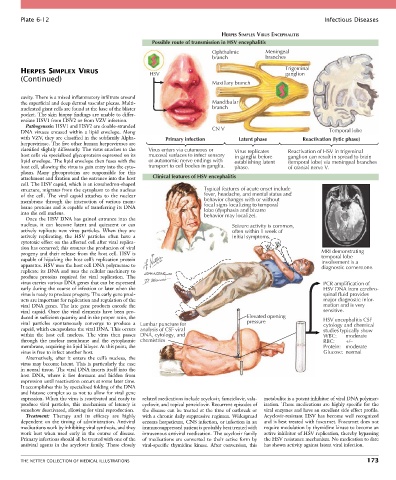
Plate 6-12                                                                                               Infectious Diseases

                                                                              HERPES SIMPLEX VIRUS ENCEPHALITIS
                                                      Possible route of transmission in HSV encephalitis
                                                                          Ophthalmic         Meningeal
                                                                          branch             branches

        HERPES SIMPLEX VIRUS                         HSV                                            Trigeminal
                                                                                                    ganglion
        (Continued)
                                                                          Maxillary branch
        cavity. There is a mixed inflammatory infiltrate around
        the superficial and deep dermal vascular plexus. Multi-           Mandibular
        nucleated giant cells are found at the base of the blister        branch
        pocket. The skin biopsy findings are unable to differ-
        entiate HSV1 from HSV2 or from VZV infection.
          Pathogenesis: HSV1 and HSV2 are double-stranded
        DNA  viruses  encased  within  a  lipid  envelope.  Along         CN V                                     Temporal lobe
        with VZV, they are classified in the subfamily Alpha-  Primary infection   Latent phase           Reactivation (lytic phase)
        herpesvirinae. The five other human herpesviruses are
        classified slightly differently. The virus attaches to the   Virus enters via cutaneous or  Virus replicates  Reactivation of HSV in trigeminal
        host cells via specialized glycoproteins expressed on its   mucosal surfaces to infect sensory  in ganglia before  ganglion can result in spread to brain
        lipid envelope. The lipid envelope then fuses with the   or autonomic nerve endings with  establishing latent  (temporal lobe) via meningeal branches
        host cell, allowing the virus to gain entry into the cyto-  transport to cell bodies in ganglia.  phase.  of cranial nerve V.
        plasm.  Many  glycoproteins  are  responsible  for  this
        attachment and fixation and the entrance into the host   Clinical features of HSV encephalitis
        cell. The HSV capsid, which is an icosahedron-shaped
        structure, migrates from the cytoplasm to the nucleus          Typical features of acute onset include
        of  the  cell.  The  viral  capsid  attaches  to  the  nuclear   fever, headache, and mental status and
        membrane  through  the  interaction  of  various  mem-         behavior changes with or without
        brane proteins and is capable of transferring its DNA          focal signs localizing to temporal
                                                                       lobe (dysphasia and bizarre
        into the cell nucleus.                                         behavior may localize).
          Once the HSV DNA has gained entrance into the
        nucleus,  it  can  become  latent  and  quiescent  or  can               Seizure activity is common,
        actively  replicate  new  virus  particles.  When  they  are             often within 1 week of
        actively  replicating,  the  HSV  particles  often  have  a              initial symptoms.
        cytotoxic effect on the affected cell after viral replica-
        tion has occurred; this ensures the production of viral
        progeny and their release from the host cell. HSV is                                                    MRI demonstrating
                                                                                                                temporal lobe
        capable of hijacking the host cell’s replication protein                                                involvement is a
        apparatus. HSV uses the host cell DNA polymerase to                                                     diagnostic cornerstone.
        replicate its DNA and uses the cellular machinery to
        produce  proteins  required  for  viral  replication.  The
        virus carries various DNA genes that can be expressed                                                    PCR amplification of
        early during the course of infection or later when the                                                   HSV DNA from cerebro-
        virus is ready to produce progeny. The early gene prod-                                                  spinal fluid provides
        ucts are important for replication and regulation of the                                                 major diagnostic infor-
        viral DNA genes. The late gene products encode the                                                       mation and is very
        viral  capsid.  Once  the  viral  elements  have  been  pro-                                             sensitive.
        duced in sufficient quantity and in the proper ratio, the                     Elevated opening           HSV encephalitis CSF
        viral  particles  spontaneously  converge  to  produce  a  Lumbar puncture for  pressure                 cytology and chemical
        capsid, which encapsulates the viral DNA. This occurs  analysis of CSF viral                             studies typically show
        within  the  host  cell  nucleus.  The  virus  then  passes  DNA, cytology, and                          WBC:    moderate
        through  the  nuclear  membrane  and  the  cytoplasmic  chemistries                                      RBC:    +/–
        membrane, acquiring its lipid bilayer. At this point, the                                                Protein:   moderate
        virus is free to infect another host.                                                                    Glucose:  normal
          Alternatively,  after  it  enters  the  cell’s  nucleus,  the
        virus may become latent. This is particularly the case
        in neural tissue. The viral DNA inserts itself into the
        host  DNA,  where  it  lies  dormant  and  hidden  from
        expression until reactivation occurs at some later time.
        It accomplishes this by specialized folding of the DNA
        and histone complex so as not to allow for viral gene
        expression. When the virus is reactivated and ready to   related medications include acyclovir, famciclovir, vala-  metabolite is a potent inhibitor of viral DNA polymer-
        produce  viral  particles,  this  mechanism  of  latency  is   cyclovir, and topical penciclovir. Recurrent episodes of   ization. These medications are highly specific for the
        somehow deactivated, allowing for viral reproduction.  the disease can be treated at the time of outbreak or   viral enzymes and have an excellent side effect profile.
          Treatment:  Therapy  and  its  efficacy  are  highly   with a chronic daily suppressive regimen. Widespread   Acyclovir-resistant  HSV  has  become  well  recognized
        dependent  on  the  timing  of  administration.  Antiviral   eczema herpeticum, CNS infection, or infection in an   and is best treated with foscarnet. Foscarnet does not
        medications work by inhibiting viral synthesis, and they   immunosuppressed patient is probably best treated with   require modulation by thymidine kinase to become an
        work  best  when  used  early  in  the  course  of  disease.   intravenous antiviral medication. The acyclovir family   active inhibitor of HSV replication, thereby bypassing
        Primary infections should all be treated with one of the   of  medications  are  converted  to  their  active  form  by   the HSV resistance mechanism. No medication to date
        antiviral  agents  in  the  acyclovir  family.  These  closely   viral-specific  thymidine  kinase.  After  conversion,  this   has shown activity against latent viral infection.


        THE NETTER COLLECTION OF MEDICAL ILLUSTRATIONS                                                                          173