
Page 286 - ACCCN's Critical Care Nursing
P. 286
Cardiac Rhythm Assessment and Management 263
FIGURE 11.24 Sinus rhythm at 65/min before onset of ventricular tachycardia. Note a ventricular ectopic emerges from the T wave of the third sinus beat
(R-on-T ventricular ectopic), precipitating ventricular tachycardia (VT). The VT is then sustained at a regular rate of 220/min, with the characteristic wide
QRS and ST/T in opposite direction to the QRS.
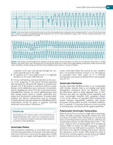
FIGURE 11.25 Probable ventricular flutter. The complexes are broad, regular and monomorphic (one shape), but it is difficult to know which is the QRS
deflection and which is the T wave. This feature plus the very fast rate of 300/min or more are the typical defining characteristics of this uncommon but
serious arrhythmia, recorded during recovery from tricyclic antidepressant overdose in a 16-year-old female.
originates at the apex and spreads through the ven- waves, ventricular flutter has earned its own classifica-
tricles upwards and to the right. tion. An example is shown in Figure 11.25. The diagnos-
32
● ST segment and T wave displacement is in opposite tic separation from other types of VT is clinically
direction to the major QRS direction. unimportant, and treatment should follow normal guide-
lines for VT.
If VT is not self-limiting, treatment depends on the sever-
ity of the symptoms. If the patient becomes pulseless
and unconscious, advanced life support is initiated (see Ventricular Fibrillation
Chapter 24). If the patient is conscious and has a pulse, During ventricular fibrillation there is no recognisable
therapy can be undertaken more cautiously. Occasionally, QRS complex. Instead, there is an irregular and wholly
5,9
robust coughing may revert VT in the cooperative patient. disorganised undulation about the baseline. There
Antiarrhythmic therapy (at slower administration rates are deflections, which at times approach rates of 300–
than during cardiac arrest) is usually undertaken first, 500/min, but these are typically of low amplitude and
along with biochemical normalisation. If unsuccessful, none convincingly resemble QRS complexes (Figure
sedation and elective cardioversion may be necessary. 11.26). In the absence of organised QRS complexes the
Consideration for internal cardioverter defibrillator (ICD) patient becomes immediately pulseless, and uncon-
implantation should be given to patients surviving sciousness follows within seconds. Immediate defibrilla-
ventricular tachycardia or fibrillation. 34,35 tion is required. If VF persists treatment occurs according
to standing basic and advanced life support guidelines.
Practice tip Polymorphic Ventricular Tachycardias
These forms of VT do not have a single QRS morphology.
Initial tolerance of VT may be evident, only to be followed by Rather, the QRS complexes during the rhythm vary from
abrupt deterioration when reserves or compensatory mecha- one shape to another, either alternating on a beat-to-beat
nisms are exhausted. Emergency responses should always be basis or switching between groups of beats, with first one
activated on initial identification. 9,32
morphology and then another (bidirectional VT). The
more common form of polymorphic VT is Torsades de
Pointes (TdP), in which the QRS undergoes a gradual
Ventricular Flutter transition from one QRS pattern to another. The descrip-
This uncommon arrhythmia is most likely just a subset tive French term, literally ‘twisting of the points’, refers to
of ventricular tachycardia, but because of its rapid rate (at the appearance of the ‘points’ (QRS direction), which is
times up to 300/min or more) and the appearance of QRS first positive and then negative, usually with an ill-defined
complexes that are largely indistinguishable from the T transition between the two (Figure 11.27). 28,36,37