
Page 287 - ACCCN's Critical Care Nursing
P. 287
264 P R I N C I P L E S A N D P R A C T I C E O F C R I T I C A L C A R E
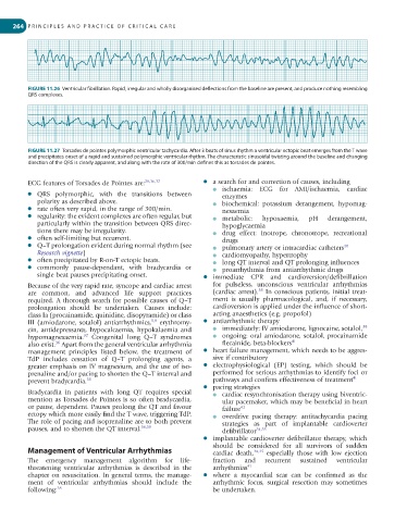
FIGURE 11.26 Ventricular fibrillation. Rapid, irregular and wholly disorganised deflections from the baseline are present, and produce nothing resembling
QRS complexes.
FIGURE 11.27 Torsades de pointes polymorphic ventricular tachycardia. After 3 beats of sinus rhythm a ventricular ectopic beat emerges from the T wave
and precipitates onset of a rapid and sustained polymorphic ventricular rhythm. The characteristic sinusoidal twisting around the baseline and changing
direction of the QRS is clearly apparent, and along with the rate of 300/min defines this as torsades de pointes.
ECG features of Torsades de Pointes are: 28,36,37 ● a search for and correction of causes, including
● ischaemia: ECG for AMI/ischaemia, cardiac
● QRS polymorphic, with the transitions between enzymes
polarity as described above. ● biochemical: potassium derangement, hypomag-
● rate often very rapid, in the range of 300/min. nesaemia
● regularity: the evident complexes are often regular, but ● metabolic: hypoxaemia, pH derangement,
particularly within the transition between QRS direc- hypoglycaemia
tions there may be irregularity. ● drug effect: inotrope, chronotrope, recreational
● often self-limiting but recurrent. drugs
● Q–T prolongation evident during normal rhythm (see ● pulmonary artery or intracardiac catheters 40
Research vignette) ● cardiomyopathy, hypertrophy
● often precipitated by R-on-T ectopic beats. ● long QT interval and QT prolonging influences
● commonly pause-dependant, with bradycardia or ● proarrhythmia from antiarrhythmic drugs
single beat pauses precipitating onset. ● immediate CPR and cardioversion/defibrillation
Because of the very rapid rate, syncope and cardiac arrest for pulseless, unconscious ventricular arrhythmias
38
are common, and advanced life support practices (cardiac arrest). In conscious patients, initial treat-
required. A thorough search for possible causes of Q–T ment is usually pharmacological, and, if necessary,
prolongation should be undertaken. Causes include: cardioversion is applied under the influence of short-
class Ia (procainamide, quinidine, disopyramide) or class acting anaesthetics (e.g. propofol)
5,9
III (amiodarone, sotalol) antiarrhythmics, erythromy- ● antiarrhythmic therapy
cin, antidepressants, hypocalcaemia, hypokalaemia and ● immediately: IV amiodarone, lignocaine, sotalol, 38
32
hypomagnesaemia. Congenital long Q–T syndromes ● ongoing: oral amiodarone, sotalol, procainamide
36
also exist. Apart from the general ventricular arrhythmia flecainide, beta-blockers 41
management principles listed below, the treatment of ● heart failure management, which needs to be aggres-
TdP includes cessation of Q–T prolonging agents, a sive if contributory
greater emphasis on IV magnesium, and the use of iso- ● electrophysiological (EP) testing, which should be
prenaline and/or pacing to shorten the Q–T interval and performed for serious arrhythmias to identify foci or
prevent bradycardia. 38 pathways and confirm effectiveness of treatment 41
● pacing strategies
Bradycardia in patients with long QT requires special ● cardiac resynchronisation therapy using biventric-
mention as Torsades de Pointes is so often bradycardia, ular pacemaker, which may be beneficial in heart
or pause, dependent. Pauses prolong the QT and favour failure 42
ectopy which more easily find the T wave, triggering TdP. ● overdrive pacing therapy: antitachycardia pacing
The role of pacing and isoprenaline are to both prevent strategies as part of implantable cardioverter
pauses, and to shorten the QT interval. 36,39 defibrillator 34,35
● implantable cardioverter defibrillator therapy, which
should be considered for all survivors of sudden
Management of Ventricular Arrhythmias cardiac death, 34,35 especially those with low ejection
The emergency management algorithm for life- fraction and recurrent sustained ventricular
threatening ventricular arrhythmias is described in the arrhythmias 41
chapter on resuscitation. In general terms, the manage- ● where a myocardial scar can be confirmed as the
ment of ventricular arrhythmias should include the arrhythmic focus, surgical resection may sometimes
following: 38 be undertaken.