
Page 288 - ACCCN's Critical Care Nursing
P. 288
Cardiac Rhythm Assessment and Management 265
other class III drugs (e.g. sotalol) and class IA agents,
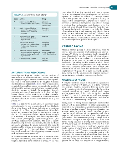
TABLE 11.1 Antiarrhythmic classifications 43 there is a risk of Q–T interval prolongation and the devel-
opment of Torsades de Pointes. 41,48 Although sotalol
Class Action Drugs carries the greatest risk of this arrhythmia, it may be
selected when amiodarone side effects need to be avoided,
IA Sodium channel blockers: quinidine
action potential prolongation procainamide or when combined antiarrhythmic–beta-blocker therapy
disopyramide is desired, (e.g. arrhythmias postinfarction or in the
setting of heart failure). Lignocaine, the front-line ven-
IB Sodium channel blockers: lignocaine
accelerate repolarisation; mexiletine tricular antiarrhythmic for many years, lacks the efficacy
shorten action potential of amiodarone, but is well tolerated and effective in the
49
duration setting of the ischaemic myocardium. Whatever the
IC Potent sodium channel flecainide choice of antiarrhythmic, additional attention should
blockers: little effect on always be directed to biochemical correction, in particu-
repolarisation lar serum magnesium, potassium and pH. 38
II Beta-blockers: depress metoprolol
automaticity (prolong phase propanolol
4); indirect prolongation esmolol CARDIAC PACING
phase 2
Artificial cardiac pacing is most commonly used to
III Potassium (outward) channel amiodarone
blockers: prolong duration of sotalol (beta-blocker provide protection against bradycardia and/or atrioven-
action potential (prolonged with class II actions) tricular (AV) block. Slow heart rates can be sustained at
repolarisation) more physiological rates by repetitive electrical stimula-
IV Calcium channel blockers verapamil tion, delivered by a pacemaker at a programmed rate.
diltiazem Temporary pacing may be provided as an emergency
intervention, providing rhythm protection whilst revers-
ible factors are overcome (biochemical or drug influence,
myocardial ischaemia or infarction) or as support until
confirmation of the need for permanent pacemaker
50
implantation. Separate from such bradycardia protec-
ANTIARRHYTHMIC MEDICATIONS tion, pacing may be undertaken to improve haemody-
Antiarrhythmic drugs are classified partly on the basis of namic status, or to treat or suppress arrhythmias.
beta-receptor or membrane channel activity, and partly
by their physiological effects on the cardiac action poten- PRINCIPLES OF PACING
tial. This is well represented by the Vaughan Williams A complete electrical circuit is achieved via a pacemaker
39
classification system (see Table 11.1). However, as action connected in series with pacing leads to (and from) the
potential abnormalities cannot be expediently identified myocardium. Electrical current is delivered to the heart
at the bedside, matching antiarrhythmic agents to cellular via the negative electrode of the circuit, whilst the positive
physiology cannot realistically be undertaken. Instead, electrode completes the electrical circuit and enables
antiarrhythmics are chosen partly on the basis of their sensing (detection) of the patient’s intrinsic cardiac
known efficacy, by their suitablity to atrial or ventricular rhythm. 51,52 Electrical impulses of sufficient strength
arrhythmias, and after consideration of side effects and stimulate the myocardium to depolarise (and then to
contraindications to known comorbidities in a given contract) at a rate selected by the operator.
patient. 41,42
Pacing leads (or pacing electrodes) may be positioned in
Table 11.2 depicts the classification of the major acute
antiarrhythmics in use in Australia and New Zealand, contact with the endocardium via transvenous access, or
along with doses, arrhythmic indications, precautions attached to the epicardium when the heart is exposed at
53
and side effects. Class I agents all slow phase 1 (depolari- the time of cardiac surgery. For epicardial pacing, two
sation) and so may slow down conduction and prolong separate leads or ‘wires’ are usually attached to each
the QRS. The subgroups of class I agents denote strength chamber paced, with one wire connected to each of the
(A = weakest, C = strongest) and affect repolarisation, negative and positive terminals of the pulse generator
with class IA (prolonging), IB (shortening) and IC (not (pacemaker). For transvenous pacing, a single lead is
affecting) repolarisation duration. The class II agents advanced to the apex of the right ventricle. These leads
(beta-blockers) depress automaticity, slowing the heart have a pacing electrode at their tip and a circumferential,
rate and prolonging the action potential. The class III or ‘ring’, sensing electrode slightly proximal to this. In an
agents notably prolong repolarisation, action potential emergency, these transvenous ventricular pacing wires
duration and the Q–T interval. Class IV agents slow can be inserted promptly and at least establish a support-
54
inward calcium channel flux, decreasing automaticity and ive ventricular rate. Temporary transvenous pacing is
prolonging the action potential. 37 almost always undertaken for ventricular pacing only.
While there are transvenous leads available for temporary
In the modern era, amiodarone ranks as the most effec- atrial pacing, they are more difficult to position, and their
tive agent in converting arrhythmias, but its use must be use is very infrequent. By contrast, in the cardiac surgical
weighed against its considerable side effects. 46,47 As with patient, where direct lead attachment is straightforward,