
Page 291 - ACCCN's Critical Care Nursing
P. 291
268 P R I N C I P L E S A N D P R A C T I C E O F C R I T I C A L C A R E
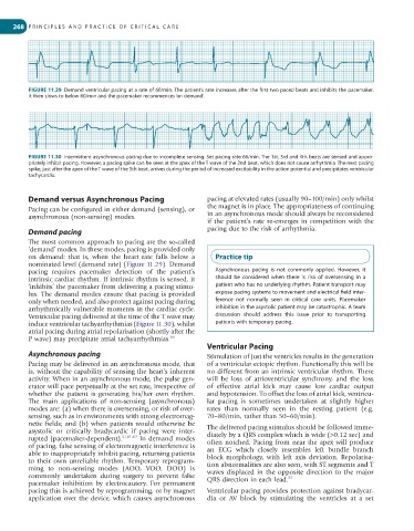
FIGURE 11.29 Demand ventricular pacing at a rate of 60/min. The patient’s rate increases after the first two paced beats and inhibits the pacemaker.
It then slows to below 60/min and the pacemaker recommences ‘on demand’.
FIGURE 11.30 Intermittent asynchronous pacing due to incomplete sensing. Set pacing rate 66/min. The 1st, 3rd and 4th beats are sensed and appro-
priately inhibit pacing. However, a pacing spike can be seen at the apex of the T wave of the 2nd beat, which does not cause arrhythmia. The next pacing
spike, just after the apex of the T wave of the 5th beat, arrives during the period of increased excitability in the action potential and precipitates ventricular
tachycardia.
Demand versus Asynchronous Pacing pacing at elevated rates (usually 90–100/min) only whilst
Pacing can be configured in either demand (sensing), or the magnet is in place. The appropriateness of continuing
asynchronous (non-sensing) modes. in an asynchronous mode should always be reconsidered
if the patient’s rate re-emerges in competition with the
Demand pacing pacing due to the risk of arrhythmia.
The most common approach to pacing are the so-called
‘demand’ modes. In these modes, pacing is provided only
on demand: that is, when the heart rate falls below a Practice tip
nominated level (demand rate) (Figure 11.29). Demand
pacing requires pacemaker detection of the patient’s Asynchronous pacing is not commonly applied. However, it
intrinsic cardiac rhythm. If intrinsic rhythm is sensed, it should be considered when there is risk of oversensing in a
‘inhibits’ the pacemaker from delivering a pacing stimu- patient who has no underlying rhythm. Patient transport may
lus. The demand modes ensure that pacing is provided expose pacing systems to movement and electrical field inter-
only when needed, and also protect against pacing during ference not normally seen in critical care units. Pacemaker
arrhythmically vulnerable moments in the cardiac cycle. inhibition in the asystolic patient may be catastrophic. A team
Ventricular pacing delivered at the time of the T wave may discussion should address this issue prior to transporting
induce ventricular tachyarrhythmias (Figure 11.30), whilst patients with temporary pacing.
atrial pacing during atrial repolarisation (shortly after the
P wave) may precipitate atrial tachyarrhythmias. 60
Ventricular Pacing
Asynchronous pacing Stimulation of just the ventricles results in the generation
Pacing may be delivered in an asynchronous mode, that of a ventricular ectopic rhythm. Functionally this will be
is, without the capability of sensing the heart’s inherent no different from an intrinsic ventricular rhythm. There
activity. When in an asynchronous mode, the pulse gen- will be loss of atrioventricular synchrony, and the loss
erator will pace perpetually at the set rate, irrespective of of effective atrial kick may cause low cardiac output
whether the patient is generating his/her own rhythm. and hypotension. To offset the loss of atrial kick, ventricu-
The main applications of non-sensing (asynchronous) lar pacing is sometimes undertaken at slightly higher
modes are: (a) when there is oversensing, or risk of over- rates than normally seen in the resting patient (e.g.
sensing, such as in environments with strong electromag- 70–80/min, rather than 50–60/min).
netic fields; and (b) when patients would otherwise be The delivered pacing stimulus should be followed imme-
asystolic or critically bradycardic if pacing were inter- diately by a QRS complex which is wide (>0.12 sec) and
rupted (pacemaker-dependent). 51,61,62 In demand modes often notched. Pacing from near the apex will produce
of pacing, false sensing of electromagnetic interference is an ECG which closely resembles left bundle branch
able to inappropriately inhibit pacing, returning patients block morphology, with left axis deviation. Repolarisa-
to their own unreliable rhythm. Temporary reprogram- tion abnormalities are also seen, with ST segments and T
ming to non-sensing modes (AOO, VOO, DOO) is waves displaced in the opposite direction to the major
commonly undertaken during surgery to prevent false QRS direction in each lead. 57
pacemaker inhibition by electrocautery. For permanent
pacing this is achieved by reprogramming, or by magnet Ventricular pacing provides protection against bradycar-
application over the device, which causes asynchronous dia or AV block by stimulating the ventricles at a set