
Page 292 - ACCCN's Critical Care Nursing
P. 292
Cardiac Rhythm Assessment and Management 269
FIGURE 11.31 Onset of ventricular pacing. At the start of the strip the patient’s heart rate is around 70/min. The pacemaker is then turned on with the
rate set at 80/min. Capture is achieved immediately, and because the pacing rate is faster there is suppression of the patient’s own rhythm. Note the wide
QRS and ST elevation during pacing. This is the expected appearance.
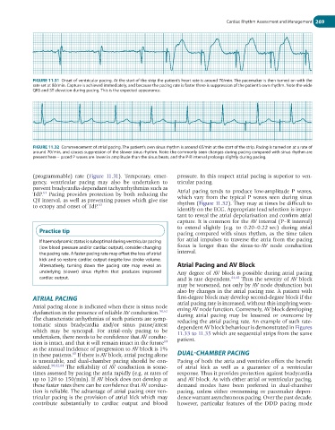
FIGURE 11.32 Commencement of atrial pacing. The patient’s own sinus rhythm is around 65/min at the start of the strip. Pacing is turned on at a rate of
around 70/min, and causes suppression of the slower sinus rhythm. Note: the commonly seen changes during pacing compared with sinus rhythm are
present here – paced P waves are lower in amplitude than the sinus beats, and the P-R interval prolongs slightly during pacing.
(programmable) rate (Figure 11.31). Temporary, emer- pressure. In this respect atrial pacing is superior to ven-
gency, ventricular pacing may also be undertaken to tricular pacing.
prevent bradycardia-dependant tachyarrhythmias such as Atrial pacing tends to produce low-amplitude P waves,
63
TdP. Pacing provides protection by both reducing the which vary from the typical P waves seen during sinus
QT interval, as well as preventing pauses which give rise rhythm (Figure 11.32). They may at times be difficult to
to ectopy and onset of TdP. 63
identify on the ECG. Appropriate lead selection is impor-
tant to reveal the atrial depolarisation and confirm atrial
capture. It is common for the AV interval (P–R interval)
to extend slightly (e.g. to 0.20–0.22 sec) during atrial
Practice tip pacing compared with sinus rhythm, as the time taken
If haemodynamic status is suboptimal during ventricular pacing for atrial impulses to traverse the atria from the pacing
(low blood pressure and/or cardiac output), consider changing focus is longer than the sinus-to-AV node conduction
the pacing rate. A faster pacing rate may offset the loss of atrial interval.
kick and so restore cardiac output despite low stroke volume.
Alternatively, turning down the pacing rate may reveal an Atrial Pacing and AV Block
underlying (slower) sinus rhythm that produces improved Any degree of AV block is possible during atrial pacing
cardiac output. and is rate dependent. 64,65 Thus the severity of AV block
may be worsened, not only by AV node dysfunction but
also by changes in the atrial pacing rate. A patient with
ATRIAL PACING first-degree block may develop second-degree block if the
atrial pacing rate is increased, without this implying wors-
Atrial pacing alone is indicated when there is sinus node ening AV node function. Conversely, AV block developing
dysfunction in the presence of reliable AV conduction. 50,62 during atrial pacing may be lessened or overcome by
The characteristic arrhythmias of such patients are symp- reducing the atrial pacing rate. An example of such rate-
tomatic sinus bradycardia and/or sinus pause/arrest dependent AV block behaviour is demonstrated in Figures
which may be syncopal. For atrial-only pacing to be 11.33 to 11.35 which are sequential strips from the same
undertaken, there needs to be confidence that AV conduc- patient.
64
tion is intact, and that it will remain intact in the future
as the annual incidence of progression to AV block is 1%
65
in these patients. If there is AV block, atrial pacing alone DUAL-CHAMBER PACING
is unsuitable, and dual-chamber pacing should be con- Pacing of both the atria and ventricles offers the benefit
sidered. 50,62,64 The reliability of AV conduction is some- of atrial kick as well as a guarantee of a ventricular
times assessed by pacing the atria rapidly (e.g. at rates of response. Thus it provides protection against bradycardia
up to 120 to 150/min). If AV block does not develop at and AV block. As with either atrial or ventricular pacing,
these faster rates there can be confidence that AV conduc- demand modes have been preferred in dual-chamber
tion is reliable. The advantage of atrial pacing over ven- pacing, unless either oversensing or pacemaker depen-
tricular pacing is the provision of atrial kick which may dence warrant asynchronous pacing. Over the past decade,
contribute substantially to cardiac output and blood however, particular features of the DDD pacing mode