
Page 295 - ACCCN's Critical Care Nursing
P. 295
272 P R I N C I P L E S A N D P R A C T I C E O F C R I T I C A L C A R E
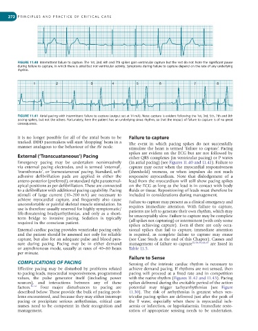
FIGURE 11.40 Intermittent failure to capture. The 1st, 2nd, 6th and 7th spikes gain ventricular capture but the rest do not. Note the significant pause
during failure to capture, in which there is atrial but not ventricular activity. Symptoms during failure to capture depend on the rate of any underlying
rhythm.
1 2 3 4 5 6 7 8
FIGURE 11.41 Atrial pacing with intermittent failure to capture (output set at 14 mA). Note: capture is evident following the 1st, 3rd, 5th, 7th and 8th
pacing spikes, but not the others. Fortunately, here the patient has an underlying sinus rhythm, so that the impact of failure to capture is of no great
consequence.
it is no longer possible for all of the atrial beats to be Failure to capture
tracked. DDD pacemakers will start ‘dropping’ beats in a The event in which pacing spikes do not successfully
manner analagous to the behaviour of the AV node. stimulate the heart is termed ‘failure to capture’. Pacing
spikes are evident on the ECG but are not followed by
External (‘Transcuataneous’) Pacing either QRS complexes (in ventricular pacing) or P waves
Emergency pacing may be undertaken noninvasively (in atrial pacing) (see Figures 11.40 and 11.41). Failure to
via external pacing electrodes, and is termed ‘external’, capture may occur when the myocardial responsiveness
‘transthoracic’, or ‘transcutaneous’ pacing. Standard, self- (threshold) worsens, or when impulses do not reach
adhesive defibrillation pads are applied in either the responsive myocardium. Note that dislodgement of a
antero-posterior (preferred), or standard right parasternal- lead from the myocardium will still show pacing spikes
apical positions as per defibrillation. These are connected on the ECG as long as the lead is in contact with body
to a defibrillator with additional pacing capability. Pacing fluids or tissue. Repositioning of leads must therefore be
stimuli of large current (10–200 mA) are necessary to included in considerations during management.
achieve myocardial capture, and frequently also cause
uncomfortable or painful skeletal muscle stimulation. Its Failure to capture may present as a clinical emergency and
use is therefore usually reserved for highly symptomatic/ requires immediate attention. With failure to capture,
life-threatening bradyarrhythmias, and only as a short- patients are left to generate their own rhythm, which may
term bridge to invasive pacing. Sedation is typically be unacceptably slow. Failure to capture may be complete
required in the conscious patient. (all spikes not capturing) or intermittent (with only some
spikes achieving capture). Even if there are only occa-
External cardiac pacing provides ventricular pacing only, sional spikes that fail to capture, immediate attention
and the patient should be assessed not only for reliable is required, as complete failure to capture may ensue
capture, but also for an adequate pulse and blood pres- (see Case Study at the end of this Chapter). Causes and
sure during pacing. Pacing may be in either demand management of failure to capture 51,58,59,68,69 are listed in
or asynchronous mode, usually at rates of 40–80 beats Table 11.5.
per minute.
Failure to Sense
COMPLICATIONS OF PACING Sensing of the intrinsic cardiac rhythm is necessary to
Effective pacing may be disturbed by problems related achieve demand pacing. If rhythms are not sensed, then
to pacing leads, myocardial responsiveness, programmed pacing will proceed at a fixed rate and in competition
values, the pulse generator itself (including power with the native rhythm (Figures 11.42 and 11.43). Pacing
sources), and interactions between any of these spikes delivered during the excitable period of the action
factors. 56-61 Four major disturbances to pacing are potential may trigger tachyarrhythmias (see Figure
described below. These provide the bulk of pacing prob- 11.30). The risk of arrhythmias is greatest when ven-
lems encountered, and because they may either interrupt tricular pacing spikes are delivered just after the peak of
pacing or precipitate serious arrhythmias, critical care the T wave, especially when there is myocardial isch-
nurses need to be competent in their recognition and aemia or infarction, or hypokalaemia. Immediate resto-
management. ration of appropriate sensing needs to be undertaken.