
Page 913 - Hematology_ Basic Principles and Practice ( PDFDrive )
P. 913
796 Part VII Hematologic Malignancies
genes, indicating the importance of mutations residing outside the
t(3;3)(q21;q26)
coding exome.
A monosomal karyotype (MK), a new cytogenetic category, is
defined as a karyotype showing two or more distinct autosomal
chromosome monosomies or one single autosomal monosomy
(excluding isolated loss of X or Y) in the presence of a structural
abnormality (see Fig. 56.20, bottom row). Initial reports indicated that
the MK in MDS, with or without monosomy for chromosomes 5
and 7, was prognostically worse than other complex karyotypes,
although later reports could not confirm these results. The reason
may be that treatment with azacitidine may have reduced the negative
der(3)del(3)(q11.2q13)inv(3)(p21q21.1) impact of MK in high-risk patients with MDS.
A B The clinical significance of loss of the Y chromosome, observed
in 10% of patients with MDS and approximately 7% of older adult
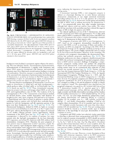
Fig. 56.23 CHROMOSOME 3 ABNORMALITIES IN MYELODYS- males without MDS, is undefined. Older adult males with MDS and
PLASTIC SYNDROME (MDS). (A) A partial karyotype from a patient with loss of Y chromosome who achieve complete hematologic remission
MDS showing a recurrent t(3;3)(q21;q26). In this rearrangement metaphase regain the Y chromosome in their marrow cells.
FISH showed on the left, RPN1 (green) and a part of MECOM (red) gene on The prognosis of patients with MDS is very heterogeneous. In
3q21 giving an impression of a “yellow” signal because of their proximity on 1997, based on the cytogenetic abnormalities identified in 816
a metaphase chromosome. The right chromosome 3 shows RPN1 (green) at patients with MDS, as well as percentage of blasts and number of
3q21, part of RPN1 (green) and MECOM (red) on 3q26 as well as unrear- cytopenia, an IPSS was proposed. According to the IPSS, 86% of
9
ranged MECOM translocated from the left chromosome 3 homolog. (B) An all cytogenetic findings can be explicitly classified according to their
unusual chromosome 3 rearrangement in MDS showing a deletion of prognostic impact. The system is highly reproducible and very simple
3q11–q13 region, followed by inversion with two breakpoints at p21 and to use, but it has certain limitations. Moreover, in the remaining 14%
q21.2. All three genes RPN1 (green), MECOM (red), and BCL6 (yellow) of patients with cytogenetic abnormalities the chromosomal abnor-
remained in their normal loci. malities had unknown prognostic significance (Fig. 56.25). This
limitation underscores two major cytogenetic classification problems
in MDS: the profound heterogeneity of acquired cytogenetic aberra-
tions in MDS and the associated challenge of designing a compre-
breakpoints were localized in pericentric regions whereas the remain- hensive cytogenetic scoring system that predicts the prognostic
ing 19% were telomeric fusions. Decondensation of pericentromeric impact of rare abnormalities. A new and comprehensive cytogenetic
heterochromatin of chromosome 1 together with centromere and scoring system based on an international data collection of 2902
repeat DNA sequences interspersed with histones and acetylated patients was recently proposed (see Fig. 56.20). Patients included
sequences may favor illegitimate recombinations leading to jumping were from the German-Austrian MDS Study Group (n = 1193), the
1q translocations. Moreover, exposure to azacitidine has been shown International MDS Risk Analysis Workshop (n = 816), the Spanish
to be associated with alterations of pericentromeric heterochromatin Hematological Cytogenetics Working Group (n = 849), and the
of chromosome 1, as well as an increase in Alu DNA repeats. Hypo- International Working Group on MDS Cytogenetics (n = 44) data-
menthilation of 1q12–21 pericentromeric region of chromosome 1 bases. In total, 19 cytogenetic categories were defined, providing clear
appears to be at least one aspect of copy number gains of 1q. prognostic classification for 91% of all patients. All abnormalities
The most frequent rearrangement of chromosome 3 involves two were arranged according to OS and development of AML to classify
bands on chromosome 3—band 3q21 and 3q26 simultaneously— their prognostic impact. The abnormalities were classified into five
which produces either t(3;3)(q21;q26) or inv(3)(q21q26) (see Fig. prognostic subgroups: very good (n = 81) included del(11q) and loss
56.20, fourth row and Fig. 56.23). These chromosomal rearrange- of Y chromosome (median OS, 61 months); good (n = 1809)
ments are present in de novo and therapy-related MDS, as well as in included normal karyotype, del(5q), del(12p), and del(20q) (all as
AML and megakaryoblastic crisis of CML. The incidence of the 3q single anomaly) and double abnormalities including del(5q) (median
rearrangements is 2% to 5%. Characteristic clinical features include OS 49 months); intermediate (n = 529) included del(7q), +8, i(17q)
an elevated platelet count, marked hyperplasia with dysplasia of (q10), +19, +21, any other single abnormality, independent clones,
megakaryocytes, and a poor prognosis with minimal or no response and double abnormalities not harboring del(5q) or −7/del(7q)
to chemotherapy and a short survival. In addition to similar clinico- (median OS 26 months); poor (n = 148) included inv(3)/t(3q)/
pathologic features, patients with 3q21q26 share molecular hetero- del(3q), −7, and double abnormalities including −7/del(7q) and
geneity in both the breakpoints and the expression pattern of the complex (three abnormalities; OS of 16 months); and very poor (n
genes near these breakpoints (see Fig. 56.23 and Fig. 56.24). The = 197) included complex karyotypes with more than three abnor-
chromosomal breakpoints, defined by FISH, in 3q26 are scattered malities (OS of 6 months) (see Fig. 56.20). This new scoring system
over several hundred kilobases in either the 5′ or the 3′ region of the proposed should be viewed as a dynamic model, open to further
EVI1 gene, whereas the breakpoints in the 3q21 region are restricted refinement as the knowledge in karyotypic abnormalities of MDS
to two smaller different genomic clusters approximately 100 kb continues to evolve. 10
downstream of the RPN1 gene (see Figs. 56.23 and 56.24). EVI1 Cytogenetic and FISH studies have relatively similar sensitivities
overexpression is observed in the majority of patients, but some in detecting an abnormal clone among patients with MDS. A FISH
patients with the 3q21q26 rearrangement do not have detectable test for targeted recurrent abnormalities in MDS should use probes
EVI1 expression, and at least 9% of patients with AML without 3q26 to detect numerical and structural anomalies of chromosome regions
abnormalities overexpress EVI1. Therefore the poor prognosis of 1q, 3q, 5q, 7, 7q31, 8, 11q, 12p, 13q, 17p, 20q, and 21. Occasional
these patients may be independent of EVI1 expression, despite the patients with normal karyotype show an occult neoplastic clone by
fact that extensive 3q26 breakpoint FISH mapping of both meta- FISH. On the other hand, using conventional cytogenetic studies,
phases and interphase nuclei suggests EVI1 involvement in numerous some patients exhibit a neoplastic clone that is not detected by FISH
novel sporadic and recurrent 3q26 rearrangements. A fusion transcript (see box on Genetic Testing for Myelodysplastic Syndrome and
of RPN1-EVI1 is rarely observed in patients with 3q21q26 rearrange- Fig. 56.20).
ments. Interestingly, functional genomic studies and allelic-specific Familial MDS is very rare. However, a recent description of four
analysis revealed experimentally that inv(3)/t(3q) results simultane- families with telomerase mutations, both in the RNA component
ously in haploinsufficiency of GATA2 and upregulation of EVI1 as a (TERC) and in the reverse transcriptase component (TERT), has
result of rearrangements in noncoding regulatory sequences of these raised the awareness of the pathologic role of telomerase mutations