
Page 914 - Hematology_ Basic Principles and Practice ( PDFDrive )
P. 914
Chapter 56 Conventional and Molecular Cytogenomic Basis of Hematologic Malignancies 797
8
B r(8)
A del(3p),dup(3)
(q26q22),r(3)
18 r(18)p11q21)
SYT at
18q11.2
MALT1 at
18q21.1
BCL2 at
18q21.3
C r(18) D r(21) 2 E r(11)
7
F r(11) G r(7)
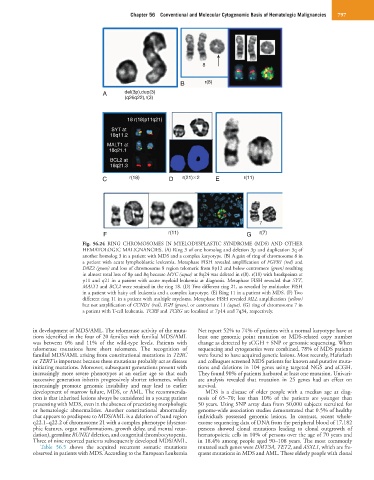
Fig. 56.24 RING CHROMOSOMES IN MYELODYSPLASTIC SYNDROME (MDS) AND OTHER
HEMATOLOGIC MALIGNANCIES. (A) Ring 3 of one homolog and deletion 3p and duplication 3q of
another homolog 3 in a patient with MDS and a complex karyotype. (B) A gain of ring of chromosome 8 in
a patient with acute lymphoblastic leukemia. Metaphase FISH revealed amplification of FGFR1 (red) and
D8Z2 (green) and loss of chromosome 8 region telomeric from 8p12 and below centromere (green) resulting
in almost total loss of 8p and 8q because MYC (aqua) at 8q24 was deleted in r(8). r(18) with breakpoints at
p11 and q21 in a patient with acute myeloid leukemia at diagnosis. Metaphase FISH revealed that SYT,
MALT1 and BCL2 were retained in the ring 18. (D) Two different ring 21, as revealed by multicolor FISH
in a patient with hairy cell leukemia and a complex karyotype. (E) Ring 11 in a patient with MDS. (F) Two
different ring 11 in a patient with multiple myeloma. Metaphase FISH revealed MLL amplification (yellow)
but not amplification of CCND1 (red), IGH (green), or centromere 11 (aqua). (G) ring of chromosome 7 in
a patient with T-cell leukemia. TCRB and TCRG are localized at 7p14 and 7q34, respectively.
in development of MDS/AML. The telomerase activity of the muta- Net report 52% to 74% of patients with a normal karyotype have at
tions identified in the four of 20 families with familial MDS/AML least one genomic point mutation or MDS-related copy number
was between 0% and 11% of the wild-type levels. Patients with change as detected by aCGH + SNP or genomic sequencing. When
telomerase mutations have short telomeres. The recognition of sequencing and cytogenetics were combined, 78% of MDS patients
familial MDS/AML arising from constitutional mutations in TERC were found to have acquired genetic lesions. Most recently, Haferlach
or TERT is important because these mutations probably act as disease and colleagues screened MDS patients for known and putative muta-
initiating mutations. Moreover, subsequent generations present with tions and deletions in 104 genes using targeted NGS and aCGH.
increasingly more severe phenotypes at an earlier age so that each They found 90% of patients harbored at least one mutation. Univari-
successive generation inherits progressively shorter telomeres, which ate analysis revealed that mutation in 25 genes had an effect on
increasingly promote genomic instability and may lead to earlier survival.
development of marrow failure, MDS, or AML. The recommenda- MDS is a disease of older people with a median age at diag-
tion is that inherited lesions always be considered in a young patient nosis of 65–70; less than 10% of the patients are younger than
presenting with MDS, even in the absence of preexisting morphologic 50 years. Using SNP array data from 50,000 subjects recruited for
or hematologic abnormalities. Another constitutional abnormality genome-wide association studies demonstrated that 0.5% of healthy
that appears to predispose to MDS/AML is a deletion of band region individuals possessed genomic lesions. In contrast, recent whole-
q22.1–q22.2 of chromosome 21 with a complex phenotype (dysmor- exome sequencing data of DNA from the peripheral blood of 17,182
phic features, organ malformations, growth delay, and mental retar- persons showed clonal mutations leading to clonal outgrowth of
dation), germline RUNX1 deletion, and congenital thrombocytopenia. hematopoietic cells in 10% of persons over the age of 70 years and
Three of nine reported patients subsequently developed MDS/AML. in 18.4% among people aged 90–108 years. The most commonly
Table 56.5 shows the acquired recurrent somatic mutations mutated such genes were DMT3A, TET2, and ASXL1, which are fre-
observed in patients with MDS. According to the European Leukemia quent mutations in MDS and AML. These elderly people with clonal