


Page 171 - Textbook of Pathology, 6th Edition
P. 171
bronchi to the other parts of the same lung or the opposite  ance is typical of tuberculous granulomas with caseation  155
           lung. This is called progressive primary tuberculosis.  necrosis.
           3. At times, bacilli may enter the circulation through erosion  Patients with HIV infection previously exposed to
           in a blood vessel and spread to various tissues and organs.  tuberculous infection have particularly high incidence of
           This is called primary miliary tuberculosis and the lesions are  reactivation of primary tuberculosis and the pattern of lesions
           seen in organs like the liver, spleen, kidney, brain and bone  in such cases is similar to that of primary tuberculosis i.e.  CHAPTER 6
           marrow.                                             with involvement of hilar lymph nodes rather than cavitary
                                                               and apical lesions in the lung. In addition, infection with M.
           4. In certain circumstances like in lowered resistance and  avium-intracellulare occurs more frequently in cases of AIDS.
           increased hypersensitivity of the host, the healed lesions of
           primary tuberculosis may get reactivated. The bacilli lying  FATE OF SECONDARY PULMONARY TUBERCULOSIS.
           dormant in acellular caseous material are activated and cause  The subapical lesions in lungs can have the following courses:
           progressive secondary tuberculosis. It affects children more  1. The lesions may  heal with fibrous scarring and
           commonly but adults may also develop this kind of   calcification.
           progression.                                        2. The lesions may coalesce together to form larger area of
                                                               tuberculous pneumonia and produce progressive secondary
           B. Secondary Tuberculosis                           pulmonary tuberculosis with the following pulmonary and
                                                               extrapulmonary involvements:                           Inflammation and Healing
           The infection of an individual who has been previously  i) Fibrocaseous tuberculosis
           infected or sensitised is called secondary, or post-primary or  ii) Tuberculous caseous pneumonia
           reinfection, or chronic tuberculosis.               iii) Miliary tuberculosis.
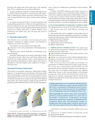
              The infection may be acquired from (Fig. 6.25):
              endogenous source such as reactivation of dormant primary  I. FIBROCASEOUS TUBERCULOSIS. The original area
           complex; or                                         of tuberculous pneumonia undergoes massive central
              exogenous source such as fresh dose of reinfection by the  caseation necrosis which may:
           tubercle bacilli.                                      either break into a bronchus from a cavity (cavitary or open
              Secondary tuberculosis occurs most commonly in lungs  fibrocaseous tuberculosis); or
           in the region of apex. Other sites and tissues which can be  remain, as a soft caseous lesion without drainage into a
           involved are tonsils, pharynx, larynx, small intestine and  bronchus or bronchiole to produce a non-cavitary lesion
           skin. Secondary tuberculosis of other organs and tissues is  (chronic fibrocaseous tuberculosis).
           described in relevant chapters later while that of lungs is  The cavity provides favourable environment for
           discussed here.                                     proliferation of tubercle bacilli due to high oxygen tension.
                                                               The cavity may communicate with bronchial tree and
           Secondary Pulmonary Tuberculosis                    becomes the source of spread of infection (‘open tuberculosis’).
                                                               The open case of secondary tuberculosis may implant
           The lesions in secondary pulmonary tuberculosis usually  tuberculous lesion on the mucosal lining of air passages
           begin as 1-2 cm apical area of consolidation of the lung, which  producing  endobronchial and endotracheal tuberculosis.
           may in time develop a small area of central caseation necrosis  Ingestion of sputum containing tubercle bacilli from
           and peripheral fibrosis. It occurs by haematogenous spread
           of infection from primary complex to the apex of the affected  endogenous pulmonary lesions may produce laryngeal and
                                                               intestinal tuberculosis.
           lung where the oxygen tension is high and favourable for
           growth of aerobic tubercle bacilli. Microscopically, the appear-
                                                                 Grossly, tuberculous cavity is spherical with thick fibrous
                                                                 wall, lined by yellowish, caseous, necrotic material and
                                                                 the lumen is traversed by thrombosed blood vessels.
                                                                 Around the wall of cavity are seen foci of consolidation.
                                                                 The overlying pleura may also be thickened (Fig. 6.26).
                                                                 Microscopically, the wall of cavity shows eosinophilic,
                                                                 granular, caseous material which may show foci of
                                                                 dystrophic calcification. Widespread coalesced
                                                                 tuberculous granulomas composed of epithelioid cells,
                                                                 Langhans’ giant cells and peripheral mantle of
                                                                 lymphocytes and having central caseation necrosis are
                                                                 seen. The outer wall of cavity shows fibrosis (Fig. 6.27).

                                                                  Complications of cavitary secondary tuberculosis are as
                                                               follows:
                                                               a) Aneurysms of patent arteries crossing the cavity
           Figure 6.25  Progressive secondary tuberculosis. A, Endogenous  producing haemoptysis.
           infection from reactivation of dormant primary complex. B, Exogenous
           infection from fresh dose of tubercle bacilli.      b) Extension to pleura producing bronchopleural fistula.

