


Page 172 - Textbook of Pathology, 6th Edition
P. 172
156


     SECTION I


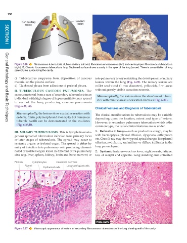
           Figure 6.26  Fibrocaseous tuberculosis. A, Non-cavitary (chronic) fibrocaseous tuberculosis (left) and cavitary/open fibrocaseous tuberculosis
           (right). B, Chronic fibrocaseous tuberculosis lung. Sectioned surface shows a cavity in the apex of the lung (arrow). There is consolidation of lung
           parenchyma surrounding the cavity.

           c) Tuberculous empyema from deposition of caseous   into pulmonary artery restricting the development of miliary
           material on the pleural surface.                    lesions within the lung (Fig. 6.29). The miliary lesions are
           d) Thickened pleura from adhesions of parietal pleura.  millet seed-sized (1 mm diameter), yellowish, firm areas
           II. TUBERCULOUS CASEOUS PNEUMONIA. The              without grossly visible caseation necrosis.
           caseous material from a case of secondary tuberculosis in an  Microscopically, the lesions show the structure of tuber-
           individual with high degree of hypersensitivity may spread  cles with minute areas of caseation necrosis (Fig. 6.30).
           to rest of the lung producing caseous pneumonia
           (Fig. 6.28, A).
                                                               Clinical Features and Diagnosis of Tuberculosis

            Microscopically, the lesions show exudative reaction with  The clinical manifestations in tuberculosis may be variable
            oedema, fibrin, polymorphs and monocytes but numerous  depending upon the location, extent and type of lesions.
            tubercle bacilli can be demonstrated in the exudates  However, in secondary pulmonary tuberculosis which is the
     General Pathology and Basic Techniques
            (Fig. 6.28,B).                                     common type, the usual clinical features are as under:
           III. MILIARY TUBERCULOSIS. This is lymphohaemato-   1. Referable to lungs—such as productive cough, may be
           genous spread of tuberculous infection from primary focus  with haemoptysis, pleural effusion, dyspnoea, orthopnoea
           or later stages of tuberculosis. The spread may occur to  etc. Chest X-ray may show typical apical changes like pleural
           systemic organs or isolated organ. The spread is either by  effusion, nodularity, and miliary or diffuse infiltrates in the
           entry of infection into pulmonary vein producing dissemi-  lung parenchyma.
           nated or isolated organ lesion in different extra-pulmonary  2. Systemic features—such as fever, night sweats, fatigue,
           sites (e.g. liver, spleen, kidney, brain and bone marrow) or  loss of weight and appetite. Long-standing and untreated


           Figure 6.27  Microscopic appearance of lesions of secondary fibrocaseous tuberculosis of the lung showing wall of the cavity.

