


Page 173 - Textbook of Pathology, 6th Edition
P. 173
157


                                                                                                                      CHAPTER 6


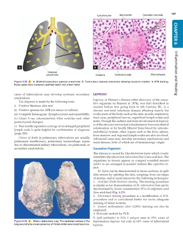
           Figure 6.28  A, Bilateral tuberculous caseous pneumonia. B, Tuberculous caseous pneumonia showing exudative reaction. In AFB staining,  Inflammation and Healing
           these cases have numerous acid-fast bacilli (not shown here).

           cases of tuberculosis may develop systemic secondary  LEPROSY
           amyloidosis.                                        Leprosy or Hansen’s disease (after discovery of the causa-
              The diagnosis is made by the following tests:    tive organism by Hansen in 1874), was first described in
           i) Positive Mantoux skin test.                      ancient Indian text going back to 6th  Century BC, is a
           ii) Positive sputum for AFB (on smear or culture).  chronic non-fatal infectious disease affecting mainly the
           iii) Complete haemogram  (lymphocytosis and raised ERR).  cooler parts of the body such as the skin, mouth, respiratory
           iv) Chest X-ray (characteristic hilar nodules and other  tract, eyes, peripheral nerves, superficial lymph nodes and
           parenchymal changes).                               testis. Though the earliest and main involvement in leprosy
           v) Fine needle aspiration cytology of an enlarged peripheral  is of the skin and nerves but in bacteraemia from endothelial
                                                               colonisation or by bacilli filtered from blood by reticulo-
           lymph node is quite helpful for confirmation of diagnosis  endothelial system, other organs such as the liver, spleen,
           (page 282).                                         bone marrow and regional lymph nodes are also involved.
              Causes of death in pulmonary tuberculosis are usually  Advanced cases may develop secondary amyloidosis and
           pulmonary insufficiency, pulmonary haemorrhage, sepsis  renal disease, both of which are of immunologic origin.
           due to disseminated miliary tuberculosis, cor pulmonale or
           secondary amyloidosis.                              Causative Organism
                                                               The disease is caused by Mycobacterium leprae which closely
                                                               resembles Mycobacterium tuberculosis but is less acid-fast. The
                                                               organisms in tissues appear as compact rounded masses
                                                               (globi) or are arranged in parallel fashion like cigarettes-in-
                                                               pack.
                                                                  M. leprae can be demonstrated in tissue sections, in split
                                                               skin smears by splitting the skin, scrapings from cut edges
                                                               of dermis, and in nasal smears by the following techniques:
                                                               1. Acid-fast (Ziehl-Neelsen) staining. The staining procedure
                                                               is similar as for demonstration of M. tuberculosis but can be
                                                               decolourised by lower concentration (5%) of sulphuric acid
                                                               (less acid-fast) (Fig. 6.31).
                                                               2. Fite-Faraco staining procedure is a modification of Z.N.
                                                               procedure and is considered better for more adequate
                                                               staining of tissue sections.
                                                               3. Gomori methenamine silver (GMS) staining can also be
                                                               employed.
                                                               4. Molecular methods by PCR.
                                                               5.  IgM antibodies to PGL-1 antigen seen in 95% cases of
           Figure 6.29  Miliary tuberculosis lung. The sectioned surface of the  lepromatous leprosy but only in 60% cases of tuberculoid
           lung parenchyma shows presence of minute millet-seed sized tubercles.  leprosy.

