


Page 174 - Textbook of Pathology, 6th Edition
P. 174
158


     SECTION I


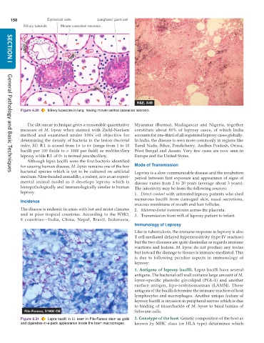
           Figure 6.30  Miliary tubercles in lung  having minute central caseation necrosis.


              The slit smear technique gives a reasonable quantitative  Myanmar (Burma), Madagascar and Nigeria, together
           measure of  M. leprae when stained with Ziehl-Neelsen  constitute about 80% of leprosy cases, of which India
           method and examined under 100x oil objective for    accounts for one-third of all registered leprosy cases globally.
           determining the density of bacteria in the lesion (bacterial  In India, the disease is seen more commonly in regions like
           index, BI). B.I. is scored from 1+ to 6+ (range from 1 to 10  Tamil Nadu, Bihar, Pondicherry, Andhra Pradesh, Orissa,
           bacilli per 100 fields to > 1000 per field) as  multibacillary  West Bengal and Assam. Very few cases are now seen in
           leprosy while B.I. of 0+ is termed paucibacillary.  Europe and the United States.
              Although lepra bacilli were the first bacteria identified
           for causing human disease, M. leprae remains one of the few  Mode of Transmission
           bacterial species which is yet to be cultured on artificial  Leprosy is a slow communicable disease and the incubation
     General Pathology and Basic Techniques
           medium. Nine-banded armadillo, a rodent, acts as an experi-  period between first exposure and appearance of signs of
           mental animal model as it develops leprosy which is  disease varies from 2 to 20 years (average about 3 years).
           histopathologically and immunologically similar to human  The infectivity may be from the following sources:
           leprosy.                                            1. Direct contact with untreated leprosy patients who shed
                                                               numerous bacilli from damaged skin, nasal secretions,
           Incidence
                                                               mucous membrane of mouth and hair follicles.
           The disease is endemic in areas with hot and moist climates  2. Materno-foetal transmission across the placenta.
           and in poor tropical countries. According to the WHO,  3. Transmission from milk of leprosy patient to infant.
           8 countries—India, China, Nepal, Brazil, Indonesia,
                                                               Immunology of Leprosy
                                                               Like in tuberculosis, the immune response in leprosy is also
                                                               T cell-mediated delayed hypersensitivity (type IV reaction)
                                                               but the two diseases are quite dissimilar as regards immune
                                                               reactions and lesions. M. leprae do not produce any toxins
                                                               but instead the damage to tissues is immune-mediated. This
                                                               is due to following peculiar aspects in immunology of
                                                               leprosy:
                                                               1. Antigens of leprosy bacilli.  Lepra bacilli have several
                                                               antigens. The bacterial cell wall contains large amount of M.
                                                               leprae-specific phenolic glycolipid (PGL-1) and another
                                                               surface antigen, lipo-arabinomannan (LAMN). These
                                                               antigens of the bacilli determine the immune reaction of host
                                                               lymphocytes and macrophages. Another unique feature of
                                                               leprosy bacilli is invasion in peripheral nerves which is due
                                                               to binding of trisaccharide of M. leprae to basal lamina of
                                                               Schwann cells.

           Figure 6.31  Lepra bacilli in LL seen in Fite-Faraco stain as globi  2. Genotype of the host. Genetic composition of the host as
           and cigarettes-in-a-pack appearance inside the foam macrophages.  known by MHC class (or HLA type) determines which

